. 2019 Apr 11;8(2):39.
doi: 10.3390/antibiotics8020039.

Rifampicin-Loaded Mesoporous Silica Nanoparticles for the Treatment of Intracellular Infections

Affiliations

Rifampicin-Loaded Mesoporous Silica Nanoparticles for the Treatment of Intracellular Infections

Santhni Subramaniam et al. Antibiotics (Basel). .

Abstract

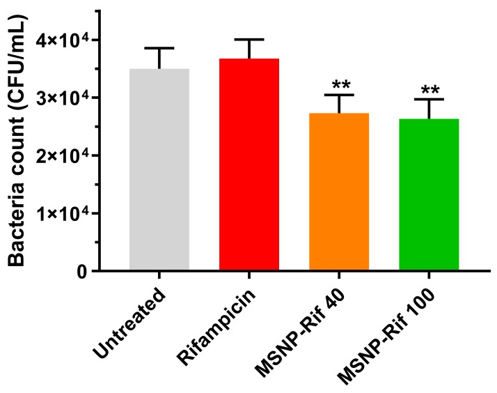
Infectious diseases remain a major burden in today's world, causing high mortality rates and significant economic losses, with >9 million deaths per year predicted by 2030. Invasion of host cells by intracellular bacteria poses treatment challenges due to the poor permeation of antimicrobials into the infected cells. To overcome these limitations, mesoporous silica nanoparticles (MSNP) loaded with the antibiotic rifampicin were investigated as a nanocarrier system for the treatment of intracellular bacterial infection with specific interest in the influence of particle size on treatment efficiency. An intracellular infection model was established using small colony variants (SCV) of S. aureus in macrophages to systemically evaluate the efficacy of rifampicin-loaded MSNP against the pathogen as compared to a rifampicin solution. As hypothesized, the superior uptake of MSNP by macrophages resulted in an enhanced treatment efficacy of the encapsulated rifampicin as compared to free antibiotic. This study provides a potential platform to improve the performance of currently available antibiotics against intracellular infections.

Keywords: MSNP; SCVs; antibiotics; intracellular infections; macrophages; nanoparticles; rifampicin.


Conflict of interest statement

The authors declare no conflict of interest.

Figures

Figure 1
Figure 1
(Left) Transmission electron microscopy (TEM) images of (A) mesoporous silica nanoparticles with particle sizes of 40 nm (MSNP-40) and (B) mesoporous silica nanoparticles with particle sizes of 100 nm (MSNP-100). (Right) Particle size distribution of MSNP-40 and MSNP-100 dispersed in Dubelcco’s modified eagle medium (DMEM) as measured by dynamic light scattering (DLS). The peak at 10 nm represents the fetal bovine serum (FBS) present in DMEM [34].
Figure 2
Figure 2
Fourier transform infrared (FTIR) spectra of MSNP-40, MSNP-100, and cetyltrimethylammonium bromide (CTAB) following solvent extraction.
Figure 3
Figure 3
Cellular viability as determined via MTT assay (mean ± SD, n = 3).
Figure 4
Figure 4
(A) Raw data obtained from fluorescence-activated cell sorter (FACS) indicating the shift in fluorescence intensity (Note: Graphs shown are only from one sample). C1 = untreated, 401 = MSNP-40, 1001 = MSNP-100. (B) Uptake of rhodamine-loaded MSNP quantified by FACS ((mean ± SD, n = 3). (p < 0.05 for MSNP-40 compared to MSNP-100). (CF) Laser scanning confocal microscopy (LCMS) images of (C,D) untreated cells, (E) MSNP-40 and (F) MSNP-100. Nuclei were stained with DAPI (Blue), MSNP were stained with rhodamine (Green) and the cell cytoskeleton was stained with wheat germ agglutinin—Alexa fluor 633 (Red).
Figure 5
Figure 5
Release kinetics of rifampicin from (left) MSNP in phosphate buffer solution (PBS) (pH 7.4) and (right) acetate buffer (pH 5.0) (mean ± SD, n = 3). Note: error bars are smaller than symbols.
Figure 6
Figure 6
The intracellular presence of small colony variants (SCV) S. aureus (stained dark blue) as observed under the light microscope using Leishman’s staining.
Figure 7
Figure 7
Efficacy of rifampicin and MSNP-Rif against SCV S. aureus (mean ± SD, n = 3). (Note: ** indicates MSNP-40 and MSNP-100 significantly different to untreated and unformulated rifampicin, p < 0.05).

References

    1. America, I.D.S.O. Antimicrobial Resistance. [(accessed on 1 March 2018)]; Available online: https://www.idsociety.org/public-health/antimicrobial-resistance/antimic...
    1. Kamaruzzaman N.F., Kendall S., Good L. Targeting the hard to reach: Challenges and novel strategies in the treatment of intracellular bacterial infections. Br. J. Pharmacol. 2017;174:2225–2236. doi: 10.1111/bph.13664. - DOI - PMC - PubMed
    1. Orfila J. Definition of intracellular pathogens. Clin. Microbiol. Infect. 1996;1:S1–S2. doi: 10.1111/j.1469-0691.1996.tb00581.x. - DOI - PubMed
    1. Hampton M.B., Kettle A.J., Winterbourn C.C. Inside the neutrophil phagosome: Oxidants, myeloperoxidase, and bacterial killing. Blood. 1998;92:3007. - PubMed
    1. Muro S. Drug Delivery across Physiological Barriers. Pan Stanford Publishing Pte. Ltd.; Singapore: 2016.