Review
. 2019 Apr 11;11(4):827.
doi: 10.3390/nu11040827.

The Role of Autophagy in Liver Epithelial Cells and Its Impact on Systemic Homeostasis

Affiliations
Review

The Role of Autophagy in Liver Epithelial Cells and Its Impact on Systemic Homeostasis

Luana Tomaipitinca et al. Nutrients. .

Abstract

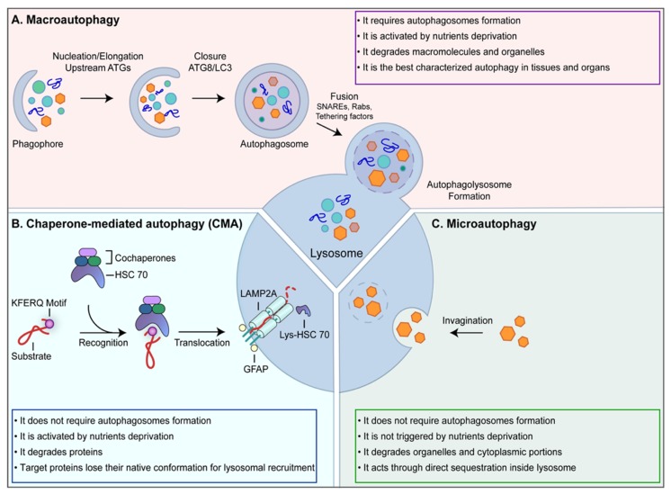
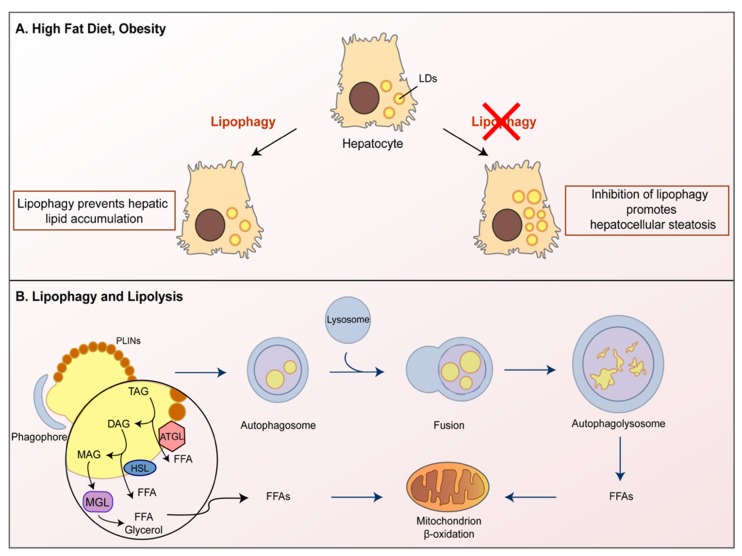
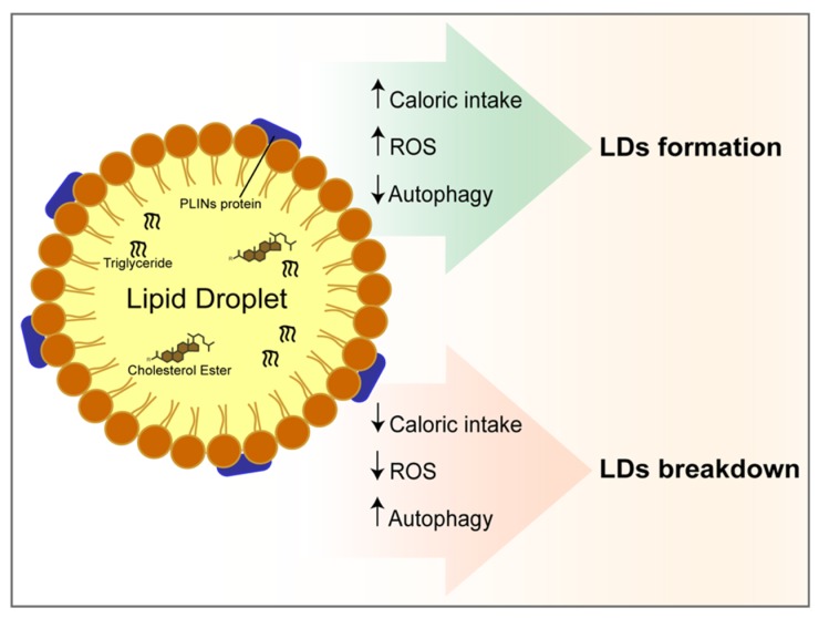
: Autophagy plays a role in several physiological and pathological processes as it controls the turnover rate of cellular components and influences cellular homeostasis. The liver plays a central role in controlling organisms' metabolism, regulating glucose storage, plasma proteins and bile synthesis and the removal of toxic substances. Liver functions are particularly sensitive to autophagy modulation. In this review we summarize studies investigating how autophagy influences the hepatic metabolism, focusing on fat accumulation and lipids turnover. We also describe how autophagy affects bile production and the scavenger function within the complex homeostasis of the liver. We underline the role of hepatic autophagy in counteracting the metabolic syndrome and the associated cardiovascular risk. Finally, we highlight recent reports demonstrating how the autophagy occurring within the liver may affect skeletal muscle homeostasis as well as different extrahepatic solid tumors, such as melanoma.

Keywords: autophagy; cancer; lipid droplets; liver; metabolism; oxidative stress.


Conflict of interest statement

All the Authors declare no conflict of interest.

Figures

Figure 1
Figure 1
Autophagy machinery. Autophagy is a quality-control process aimed at eliminating old or damaged cellular components. (A) Macroautophagy is the most common form of autophagy. It requires different sequential steps leading to the formation of autophagosomes. Following the fusion between autophagosomes and lysosome, the cargo is degraded and the resulting macromolecules are released back into the cytosol to be recycled; (B) chaperone-mediated autophagy (CMA) contributes to the cellular homeostasis by recycling amino acids upon proteins degradation and by eliminating abnormal or damaged proteins. CMA targets are recognized by this molecular machinery as they contain the KFERQ motif; (C) Microautophagy is involved in organelles and cytoplasmic portions turnover through direct sequestration inside lysosomes.
Figure 2
Figure 2
The representative area of normal hepatic tissue. Immunohistochemistry for CK-7, a specific cytokeratin of the biliary epithelium, that is stained in brown (BD). In blue the hepatocytes are evident (H), the white spaces are the sinusoids (S). In the center, a portal space is evident with the typical branches of the portal vein (PV), hepatic artery (HA) and several bile ducts (BD) cut on different planes (transverse or sagittal). OM 20x.
Figure 3
Figure 3
The autophagy involvement in lipid droplets turnover. Autophagy plays a key role in lipid droplets (LDs) metabolism in hepatocytes. (A) Autophagy breaks LDs inside lysosomes in a process termed lipophagy. Lipophagy can prevent lipid accumulation in hepatocytes, while the inhibition of lipophagy promotes LDs accumulation, resulting in hepatocellular steatosis. (B) Lipolysis is a process mediated by three lipases: ATGL, HSL and MGL. ATGL controls the crosstalk between lipolysis and autophagy as it regulates TAG turnover. ATGL promotes lipophagy to facilitate LD catabolism leading to the generation of Free Fatty Acids (FFAs), which are broken down by the β-oxidation process.
Figure 4
Figure 4
Autophagy at the crossroad between oxidative stress, lipids accumulation and cell death in a hypercaloric diet. Lipid droplets consist of a hydrophobic core of neutral lipids, surrounded by a phospholipid monolayer characterized by perilipins (PLINs) proteins. The core of LDs is composed of triglycerides and cholesterol esters. The figure shows the positive and negative correlation between reactive oxygen species (ROS), autophagy and diet with LDs formation or breakdown.
Figure 5
Figure 5
The role of autophagy on biliary epithelium differentiation and homeostasis. Autophagy is involved in the maintenance and functions of progenitory cells in the liver (HPC). It negatively correlates with biliary tree formation, it decreases in the early stages of HPC development while it increases in the late stages. HPC differentiation to cholangiocyte is controlled by the Notch signaling pathway. In cholangiocytes, the under nutrient-rich condition or absence of rapamycin, mTOR blocks autophagy resulting in an increased Notch-STAT3 cascade signaling pathway and promoting cellular differentiation. On the contrary, autophagy induction via rapamycin (an mTOR inhibitor) or nutrient deprivation attenuates the Notch signaling pathway resulting in a reduced biliary differentiation.
Figure 6
Figure 6
The role of autophagy in endothelial cells maintenance. Autophagy plays an important role in the regulation of the Sinusoidal endothelial liver cells (SEC) phenotype. In fact, under basal conditions, autophagy maintains SEC homeostasis. During liver injury, autophagy impairment increases oxidative stress and leads to liver fibrosis. On the other hand, a high autophagy rate induces caveolin-1 degradation, thus leading to SEC defenestration and liver fibrosis.

References

    1. Deter R.L., Baudhuin P., De Duve C. Participation of lysosomes in cellular autophagy induced in rat liver by glucagon. J. Cell Biol. 1967;35:C11–C16. doi: 10.1083/jcb.35.2.C11. - DOI - PMC - PubMed
    1. Tsukada M., Ohsumi Y. Isolation and characterization of autophagy-defective mutants of Saccharomyces cerevisiae. FEBS Lett. 1993;333:169–174. doi: 10.1016/0014-5793(93)80398-E. - DOI - PubMed
    1. Zientara-Rytter K., Subramani S. The Roles of Ubiquitin-Binding Protein Shuttles in the Degradative Fate of Ubiquitinated Proteins in the Ubiquitin-Proteasome System and Autophagy. Cells. 2019;8:40. doi: 10.3390/cells8010040. - DOI - PMC - PubMed
    1. Giampietri C., Petrungaro S., Cordella M., Tabolacci C., Tomaipitinca L., Facchiano A., Eramo A., Filippini A., Facchiano F., Ziparo E. Lipid Storage and Autophagy in Melanoma Cancer Cells. Int. J. Mol. Sci. 2017;18:1271. doi: 10.3390/ijms18061271. - DOI - PMC - PubMed
    1. Zhang Z., Yao Z., Chen Y., Qian L., Jiang S., Zhou J., Shao J., Chen A., Zhang F., Zheng S. Lipophagy and liver disease: New perspectives to better understanding and therapy. Biomed. Pharmacother. 2018;97:339–348. doi: 10.1016/j.biopha.2017.07.168. - DOI - PubMed

LinkOut - more resources