. 2019 May 21;50(5):1305-1316.e6.
doi: 10.1016/j.immuni.2019.03.010. Epub 2019 Apr 9.

Immunization with Components of the Viral Fusion Apparatus Elicits Antibodies That Neutralize Epstein-Barr Virus in B Cells and Epithelial Cells

Affiliations

Immunization with Components of the Viral Fusion Apparatus Elicits Antibodies That Neutralize Epstein-Barr Virus in B Cells and Epithelial Cells

Wei Bu et al. Immunity. .

Abstract

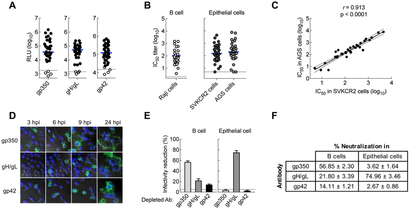
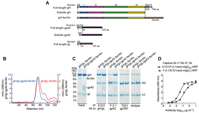
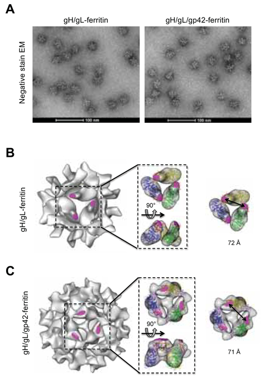
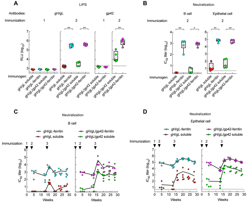
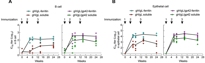
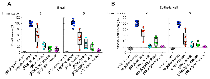
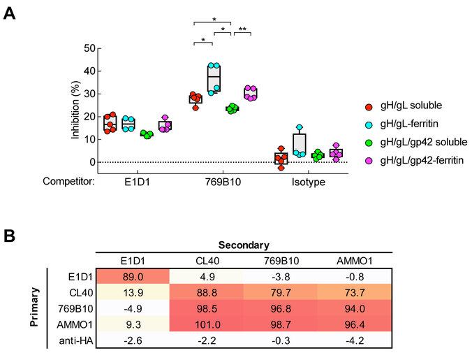
Epstein-Barr virus (EBV) causes infectious mononucleosis and is associated with epithelial-cell cancers and B cell lymphomas. An effective EBV vaccine is not available. We found that antibodies to the EBV glycoprotein gH/gL complex were the principal components in human plasma that neutralized infection of epithelial cells and that antibodies to gH/gL and gp42 contributed to B cell neutralization. Immunization of mice and nonhuman primates with nanoparticle vaccines that displayed components of the viral-fusion machinery EBV gH/gL or gH/gL/gp42 elicited antibodies that potently neutralized both epithelial-cell and B cell infection. Immune serum from nonhuman primates inhibited EBV-glycoprotein-mediated fusion of epithelial cells and B cells and targeted an epitope critical for virus-cell fusion. Therefore, unlike the leading EBV gp350 vaccine candidate, which only protects B cells from infection, these EBV nanoparticle vaccines elicit antibodies that inhibit the virus-fusion apparatus and provide cell-type-independent protection from virus infection.

Keywords: B cell lymphoma; Epstein-Barr virus; infectious mononucleosis; nanoparticle; vaccine; virus fusion.


Conflict of interest statement

DECLARATION OF INTERESTS

M.K., W.B., G.J.N. and J.I.C. are named as inventors on a patent application describing the data presented in this paper, which have been filed by the National Institutes of Health. G.N. is an employee of Sanofi.

Figures

Figure 1.
Figure 1.
Contribution of glycoprotein antibodies to B cell and epithelial cell neutralizing titers in human sera and plasma. (A) EBV gp350 antibody, gH/gL antibody, and gp42 antibody titers in human plasma measured by LIPS assay. Antibody titers are expressed as luciferase relative light units (RLU). Solid circles are samples from 34 EBV seropositive subjects (positive for viral capsid antigen [VCA] IgG antibody); open circles are the serum from EBV seronegative subjects (4 subjects). The horizontal blue line represents the median of antibody titers. The horizontal dotted lines are the cut off value defined as twice the value of the buffer control. (B) Neutralizing antibody titers in sera of EBV seropositive subjects were measured using B cell neutralization (Raji cells) and epithelial cells neutralization (SVKCR2 cells and AGS cells) assays. The horizontal dark line is the median of the neutralizing antibody titers. The dotted line represents the detection limit of the assay. (C) Neutralizing antibody titers in the two epithelial cell lines, SVKCR2 and AGS, show a signfiicant correlation (p < 0.0001). (D) Surface of vaccinia virus (VV)-infected cells expressing EBV glycoproteins gp350, gH/gL or gp42 was stained with mAbs 72A1, E1D1, or F-2–1, respectively, followed by Alexa Fluor 488 goat anti-mouse IgG antibody at 3, 6, 9, and 24 hr post infection (hpi). (E) Reduction in EBV infection of B cells (Raji cells) and epithelial cells (SVKCR2 cells) by IVIG in which antibodies to individual EBV glycoproteins were depleted using VV-infected cells expressing EBV glycoproteins. The percentage of infectivity reduction was calculated by (1-IC50-depleted/IC50-control) ×100 in which IC50-depleted is the neutralizing antibody titer of depleted IVIG and IC50-control is the neutralizing antibody titer of IVIG incubated with control VV-infected cells. (F) Contribution of gp350, gH/gL and gp42 in human plasma to B cell and epithelial cell neutralizing antibodies. Data shown are the mean + standard errors of the mean (SEM) based on 3 independent experiments. See also Figure S1.
Figure 2.
Figure 2.
Construction and characterization of gH/gL-ferritin and gH/gL/gp42-ferritin nanoparticles. (A) Schematic representation of full-length gH, soluble gH, gH-ferritin, full-length gp42, soluble gp42, and full-length gL. gH-ferritin fusion protein was generated by fusion of gH ectodomain (domains I, II, III, and IV) to the N-terminus of the ferritin. SS is the native signal sequence of the glycoprotein, SS* is the human CD5 signal sequence, TM is the transmembrane domain and CT is the cytoplasmic tail. Amino acid (aa) position of gH, gL, and gp42 is indicated. (B) Chromatograph from size-exclusion chromatography of gH/gL-ferritin and gH/gL/gp42-ferritin nanoparticles purified from the supernatants of mammalian cells transfected with gH-ferritin and gL plasmids or gH-ferritin, gL, and gp42 plasmids. Particles were affinity purified using snowdrop lectin prior to size exclusion chromatography. Transient transfection of gH-ferritin/gL and gH-ferritin/gL/gp42 plasmids, resulted in comparable amount of nanoparticle proteins (~ 2mg/L). (C) Characterization of nanoparticles by immunoprecipitation and SDS-PAGE. Bands corresponding to gH-ferritin, gp42, and gL are indicated. Purified nanoparticles were immunoprecipitated with anti-gH/gL mAb (E1D1), anti-gp42 mAb (F-2–1), anti-gp350 mAb (72A1), or isotype control antibody. HC and LC denote antibody heavy and light chains, respectively. Faint bands running slightly slower than gH-ferritin are nonspecific background bands. (D) Sandwich ELISA using mAb E1D1 or F-2–1 to capture purified gH/gL/gp42-ferritin nanoparticles and detection with mAb F-2–1 or E1D1, respectively. The dotted line represents the background. See also Figure S2 and S3.
Figure 3.
Figure 3.
Negative-stain transmission electron microscopy images and cryo-EM analysis of nanoparticles. (A) Negative-stain transmission EM of gH/gL-ferritin and gH/gL/gp42-ferritin nanoparticles. Cryo EM reconstruction models of gH/gL-ferritin (B) and gH/gL/gp42-ferritin (C) nanoparticles. Coordinates corresponding to gH/gL (PDB ID 3PHF) and gH/gL/gp42 (PDB ID 5T1D) were fitted into cryo-EM density maps. Structures of gH (blue, green, or yellow), gL (red), gp42 (purple), are shown in ribbon representation with the mAb E1D1 binding sites (magenta). The distance between two E1D1 binding sites on particles is indicated. See also Figure S4.
Figure 4.
Figure 4.
Immunogenicity of soluble and nanoparticle gH/gL and gH/gL/gp42 immunogens in mice. BALB/c mice (n=5) were immunized intramuscularly with 0.5 μg of the indicated proteins with Sigma Adjuvant System adjuvant at week 0, 3, and 14 (corresponding to injections 1, 2, and 3, respectively). (A) Antibody titers to gH/gL and gp42 in immune sera of 2 weeks after immunizations 1 and 2 were measured by LIPS assay and shown as relative light units (RLU). The horizontal dotted line is the cutoff value defined as twice the value of the buffer control. (B) EBV B cell and epithelial cell neutralizing titers in sera following two immunizations. Data are represented as box-and-whiskers plots (box indicates lower and upper quartiles with horizontal line at median and whiskers at minimum and maximum data points). (C and D) Kinetics of B cell and epithelial cell neutralizing titers in mice. Antibody titers were determined at week 5, 13, 16, 20, 24 and 28. Each dot represents an individual mouse. The dotted lines represent the limit of detection. * p < 0.05; ** p < 0.01. See also Figure S5.
Figure 5.
Figure 5.
Immunogenicity of soluble and nanoparticle gH/gL and gH/gL/gp42 immunogens in nonhuman primates. Cynomolgus macaques were immunized intramuscularly with 50 μg of soluble gH/gL (N=5), gH/gL-ferritin (N=4), soluble gH/gL/gp42 (N=5), or gH/gL/gp42-ferritin (N=5) formulated with Sigma Adjuvant System adjuvant at week 0, 4, and 12 (corresponding to injections 1, 2, and 3, respectively). (A) B cell and (B) epithelial cell neutralization titers in sera were measured at week 0, 6, 8, 14, and 24. p = 0.0179 for gH/gL-ferritin vs. soluble gH/gL, and p = 0.1111 for gH/gL/gp42-ferritin vs. soluble gH/gL/gp42 for B cell neutralizing titers at week 6. p = 0.159 for gH/gL-ferritin vs. soluble gH/gL, and p = 0.0317 for gH/gL/gp42-ferritin vs. soluble gH/gL/gp42 for epithelial cell neutraling titers at week 6. Each dot represents an individual monkey. The dotted lines represent the limit of detection. See also Figure S6.
Figure 6.
Figure 6.
Inhibition of B cell and epithelial cell fusion by serum from monkeys immunized with soluble gH/gL, gH/gL-ferritin, soluble gH/gL/gp42, or gH/gL/gp42-ferritin. Inhibition of B cell (A) and epithelial cell (B) fusion by sera from monkeys 2 weeks after the second and the third dose of vaccine. Sera from monkeys prior to immunization (negative sera) were used as a positive control, while assays using CHO-K1 cells expressing EBV gH/gL and luciferase with or without gp42, but not EBV gB, were used as a negative control for fusion. The percentage of fusion was calculated as (RLUSerum/RLUNegative Serum) × 100%. p = 0.0159 for gH/gL-ferritin vs. soluble gH/gL; p = 0.0952 for gH/gL/gp42-ferritin vs. soluble gH/gL/gp42 for epithelial cell fusion. The dotted lines represent the background of the assay. Data are represented as box-and-whiskers plots and each dot represents an individual monkey. The figure shown is representative of two independent experiments.
Figure 7.
Figure 7.
Detection of antibodies that compete with known EBV neutralizing monoclonal antibodies in immunized monkey sera. (A) Cross-competition of immune sera to gH/gL mAbs E1D1 and 769B10. The percent inhibition of monkey sera antibody to bind gH/gL by mAb E1D1, 769B10, or isotype control antibody is shown. Data are represented as box-and-whiskers plots and each dot represents an individual monkey. * p < 0.05; ** p < 0.01. (B) Table of antibody cross-competition of mAb 769B10 by mAbs E1D1, CL40, AMMO1, and anti-HA (control). Antibody competition was measured by biolayer interferometry with recombinant gH/gL protein. Biosensors immobilized with gH/gL protein were pre-saturated with antibodies (primary) and then measured binding of secondary antibodies. See also Figure S7.

References

    1. Bachmann MF, Rohrer UH, Kündig TM, Bürki K, Hengartner H, and Zinkernagel RM (1993). The influence of antigen organization on B cell responsiveness. Science. 262, 1448–1451. - PubMed
    1. Blasco R, and Moss B (1995). Selection of recombinant vaccinia viruses on the basis of plaque formation. Gene 158, 157–162. - PubMed
    1. Brochet X, Lefranc MP, and Giudicelli V (2008). IMGT/V-QUEST: the highly customized and integrated system for IG and TR standardized V-J and V-D-J sequence analysis. Nucleic Acids Res. 36. - PMC - PubMed
    1. Chen J, Sathiyamoorthy K, Zhang X, Schaller S, Perez White BE, Jardetzky TS, and Longnecker R (2018). Ephrin receptor A2 is a functional entry receptor for Epstein–Barr virus. Nat. Microbiol 1–9. - PMC - PubMed
    1. Chesnokova LS, and Hutt-Fletcher LM (2011). Fusion of Epstein-Barr virus with epithelial cells can be triggered by αvβ5 in addition to αvβ6 and αvβ8, and integrin binding triggers a conformational change in glycoproteins gHgL. J. Virol 85, 13214–13223. - PMC - PubMed

Publication types

MeSH terms