Observational Study
. 2019 Apr 12;4(34):eaav6778.
doi: 10.1126/sciimmunol.aav6778.

AIRE expression controls the peripheral selection of autoreactive B cells

Affiliations
Observational Study

AIRE expression controls the peripheral selection of autoreactive B cells

Joel Sng et al. Sci Immunol. .

Abstract

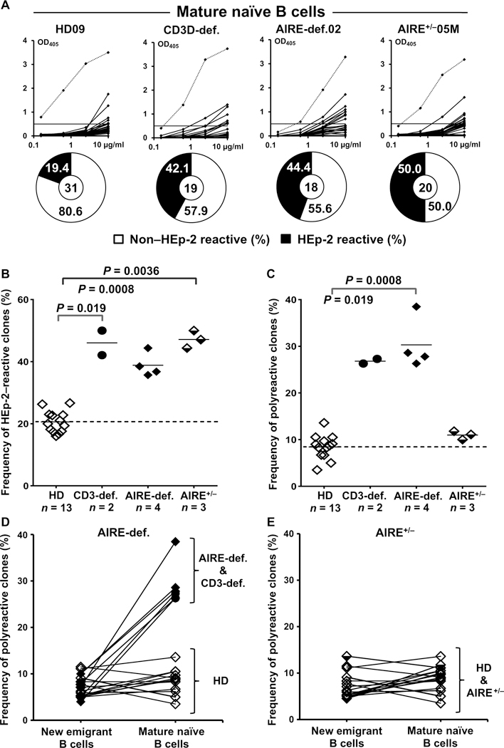
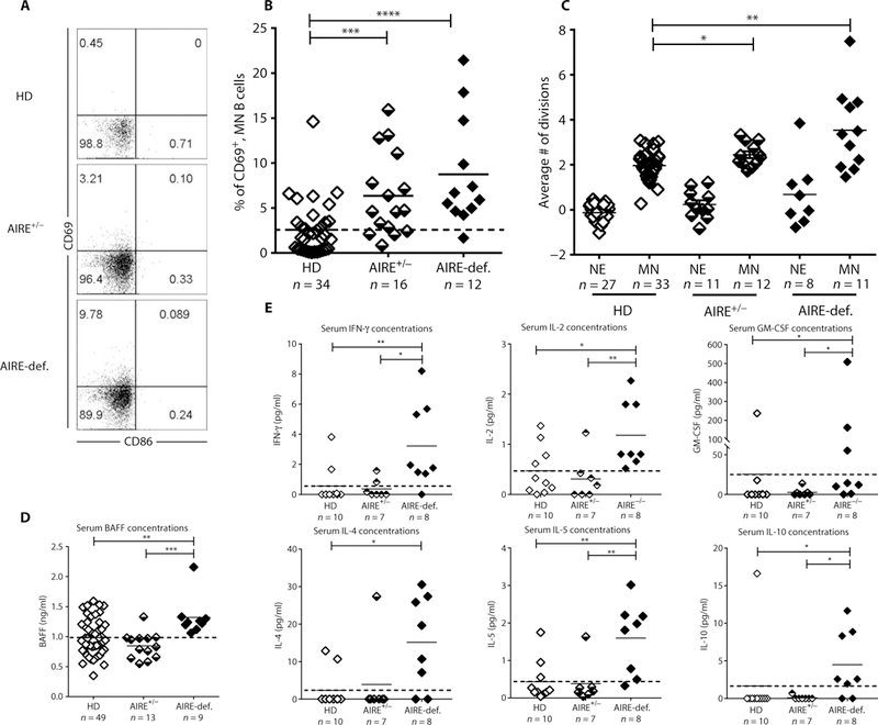
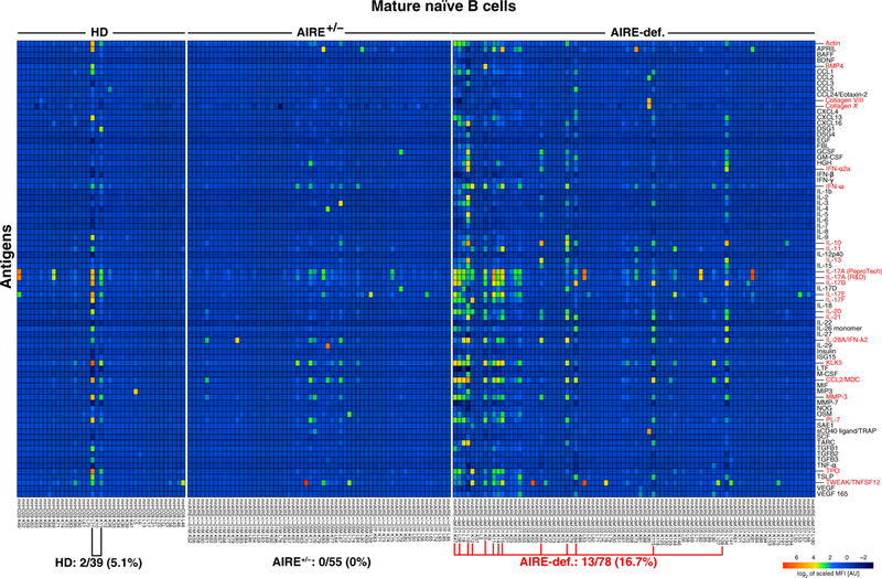
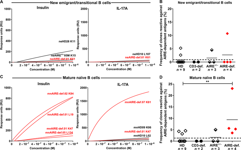
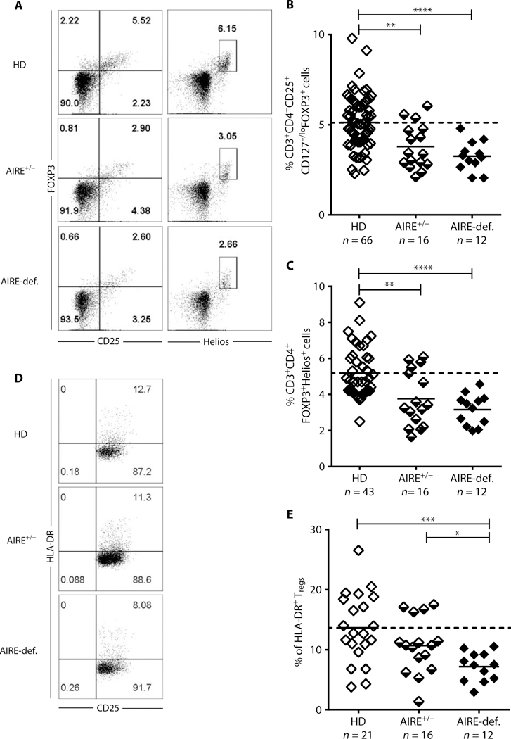
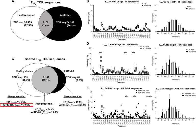
Autoimmune regulator (AIRE) mutations result in autoimmune polyendocrinopathy-candidiasis-ectodermal dystrophy (APECED) syndrome characterized by defective central T cell tolerance and the production of many autoantibodies targeting tissue-specific antigens and cytokines. By studying CD3- and AIRE-deficient patients, we found that lack of either T cells or AIRE function resulted in the peripheral accumulation of autoreactive mature naïve B cells. Proteomic arrays and Biacore affinity measurements revealed that unmutated antibodies expressed by these autoreactive naïve B cells recognized soluble molecules and cytokines including insulin, IL-17A, and IL-17F, which are AIRE-dependent thymic peripheral tissue antigens targeted by autoimmune responses in APECED. AIRE-deficient patients also displayed decreased frequencies of regulatory T cells (Tregs) that lacked common TCRβ clones found instead in their conventional T cell compartment, thereby suggesting holes in the Treg TCR repertoire of these patients. Hence, AIRE-mediated T cell/Treg selection normally prevents the expansion of autoreactive naïve B cells recognizing peripheral self-antigens.


Figures

Fig. 1.
Fig. 1.. Central B cell tolerance is functional in CD3- and AIRE-deficient patients.
(A) Antibodies from new emigrant B cells from HDs (n = 12), CD3-deficient patients (CD3-def., n = 2), AIRE-deficient patients (AIRE-def., n = 4), and AIRE+/− heterozygous relatives (AIRE+/−, n = 3) were tested by ELISA for reactivity against dsDNA, insulin, and LPS. Antibodies were considered polyreactive when they recognized all three analyzed antigens. Dotted lines show ED38-positive control. Horizontal lines show cut-off OD405 for positive reactivity. For each individual, the frequency of nonpolyreactive (open area) and polyreactive (filled area) clones is summarized in pie charts, with the total number of clones tested indicated in the centers. The frequencies of polyreactive and antinuclear new emigrant/transitional B cells are summarized in (B) and (C), respectively. Each symbol represents an individual. Solid lines show the mean, and dashed lines indicate the averaged mean value for HDs.
Fig. 2.
Fig. 2.. Defective peripheral B cell tolerance checkpoint in CD3- and AIRE-deficient patients.
(A) Antibodies from mature naïve B cells from HDs (n = 12), CD3-deficient patients (n = 2), AIRE-deficient patients (n = 4), and AIRE+/− heterozygous relatives (n = 3) were tested by ELISA for anti-HEp-2 cell reactivity. Dotted lines show ED38-positive control. Horizontal lines show cut-off OD405 for positive reactivity. For each individual, the frequency of non-HEp-2 reactive (open area) and HEp-2-reactive (filled area) clones is summarized in pie charts, with the total number of clones tested indicated in the centers. The frequencies of HEp-2-reactive and polyreactive mature naïve B cells are summarized in (B) and (C), respectively. Each symbol represents an individual. Solid lines show the mean, and dashed line indicates the averaged mean value for HDs. The frequency of polyreactive B cells and their evolution between the new emigrant/ transitional and mature naïve B cell stages in (D) CD3- and AIRE-deficient patients and (E) AIRE+/− heterozygous relatives are shown in comparison with HDs.
Fig. 3.
Fig. 3.. Mature naïve B cells from AIRE-deficient patients show increased activation and homeostatic expansion associated with elevated serum BAFF and systemic cytokine concentrations. (A)
Representative CD69 and CD86 expression on CD19+CD27CD21+CD10 mature naïve B cells from HDs, AIRE+/− heterozygous relatives, and AIRE-deficient patients. (B) Frequencies of CD69+ mature naïve B cells in indicated individuals. (C) Evaluation of the number of cell divisions undergone in vivo by KREC analysis of new emigrant (NE)/transitional and mature naïve (MN) B cells from HDs, AIRE+/− carriers, and AIRE-deficient patients. Serum concentrations of (D) BAFF and (E) IFN-γ, IL-2, GM-CSF, IL-4, IL-5, and IL-10 measured by ELISA and Luminex, respectively, in various individuals’ groups. Each symbol represents an individual in (B), (C), and (D). Solid lines show the mean, and dashed line indicates the averaged mean value for HDs. Statistically significant differences are indicated. *P ≤ 0.05, **P ≤ 0.01, ***P ≤ 0.001, ****P ≤ 0.0001.
Fig. 4.
Fig. 4.. Autoantigen array profiling of recombinant antibodies cloned from single-cell sorted mature naïve B cells from HDs, AIRE heterozygotes, and AIRE-deficient patients.
Heatmap displaying reactivity of individual mature naïve B cells from HDs (n = 2), AIRE+/− heterozygous individuals (n = 3), and AIRE-deficient patients (n = 4) against selected cytokines and other self-antigens. The frequency of self-reactive mature naïve B cells is elevated in AIRE-deficient patients compared with HDs and AIRE+/− heterozygous individuals, reflecting the accumulation of anticytokine and other autoreactive clones. MFI, mean fluorescent intensity; AU, arbitrary units.
Fig. 5.
Fig. 5.. Elevated frequency of AIRE-dependent antigen-reactive mature naïve B cells in AIRE-deficient patients.
SPR analysis of binding to AIRE-dependent antigens insulin and IL-17A by recombinant antibodies cloned from single (A) new emigrant/transitional and (C) mature naïve B cells from nine HDs, two CD3-deficient patients, three AIRE+/− heterozygous individuals, and four AIRE-deficient patients. Frequencies of new emigrant/transitional and mature naïve B cells from HDs, CD3-deficient patients, AIRE+/− heterozygous individuals, and AIRE-deficient patients reactive against AIRE-dependent antigens are represented in (B) and (D), respectively. Each symbol represents an individual. **P ≤ 0.01.
Fig. 6.
Fig. 6.. Altered Treg frequencies in AIRE-deficient patients and AIRE+/ heterozygous individuals.
(A) Representative CD25 and FOXP3 (left) and Helios and FOXP3 (right) staining on CD3+CD4+ cells from an HD, AIRE+/− heterozygous individual, and AIRE-deficient patient. (B) CD3+CD4+CD25+CD127−/loFOXP3+ and (C) CD3+CD4+FOXP3+Helios+ Treg frequencies in HDs, AIRE+/− heterozygous individuals, and AIRE-def. patients. (D) Representative CD25 and HLA-DR staining on CD3+CD4+CD25+CD127−/lo T cells and (E) CD3+CD4+CD25+CD127loHLA-DR+ T cell frequencies in HDs, AIRE+/− heterozygous individuals, and AIRE-def. patients. Each symbol represents an individual and solid lines show the mean; dashed line indicates the averaged mean value for HDs. *P ≤ 0.05, **P ≤ 0.01, ***P ≤ 0.001; **** P ≤ 0.0001.
Fig. 7.
Fig. 7.. AIRE deficiency affects the peripheral CD4+ Treg and Tconv cell TCR repertoire.
CD4+CD25hiCD127−/lo Treg and CD4+CD127+ Tconv cells were purified by fluorescence-activated cell sorting from an HD (n = 5) controls and AIRE-deficient patients (n = 5) and subjected to TCRBV sequencing. (A) Summary chart of total distinct Treg CDR3 TCR sequences obtained from pooled HD controls and AIRE-deficient patients. (B) TCRBV V gene segment usage and CDR3 length in Tregs from HDs and AIRE-deficient patients. (C) Treg TCRBV sequences common to HDs but absent in patients with APECED can be identified in the Tconv compartment of AIRE-deficient patients and HDs. Comparison of TCRBV V gene segment usage and CDR3 length in total versus shared Treg sequences in (D) HDs and (E) AIRE-deficient patients. aa, amino acids. *P ≤ 0.05, **P ≤ 0.01, ***P ≤ 0.001.

References

    1. Nagamine K, Peterson P, Scott HS, Kudoh J, Minoshima S, Heino M, Krohn KJE, Lalioti MD, Mullis PE, Antonarakis SE, Kawasaki K, Asakawa S, Ito F, Shimizu N, Positional cloning of the APECED gene. Nat. Genet. 17, 393–398 (1997). - PubMed
    1. Finnish-German APECED Consortium, An autoimmune disease, APECED, caused by mutations in a novel gene featuring two PHD-type zinc-finger domains. Nat. Genet. 17, 399–403 (1997). - PubMed
    1. Anderson MS, Venanzi ES, Klein L, Chen Z, Berzins SP, Turley SJ, von Boehmer H, Bronson R, Dierich A, Benoist C, Mathis D, Projection of an immunological self shadow within the thymus by the aire protein. Science 298, 1395–1401 (2002). - PubMed
    1. Kärner J, Meager A, Laan M, Maslovskaja J, Pihlap M, Remm A, Juronen E, Wolff ASB, Husebye ES, Podkrajsek KT, Bratanic N, Battelino T, Willcox N, Peterson P, Kisand K, Anti-cytokine autoantibodies suggest pathogenetic links with autoimmune regulator deficiency in humans and mice. Clin. Exp. Immunol. 171, 263–272 (2013). - PMC - PubMed
    1. Ramsey C, Winqvist O, Puhakka L, Halonen M, Moro A, Kämpe O, Eskelin P, Pelto-Huikko M, Peltonen L, Aire deficient mice develop multiple features of APECED phenotype and show altered immune response. Hum. Mol. Genet. 11, 397–409 (2002). - PubMed

Publication types

MeSH terms