. 2019 Apr 12;9(1):5984.
doi: 10.1038/s41598-019-42401-9.

Characterization of donor and recipient CD8+ tissue-resident memory T cells in transplant nephrectomies

Affiliations

Characterization of donor and recipient CD8+ tissue-resident memory T cells in transplant nephrectomies

Kitty de Leur et al. Sci Rep. .

Abstract

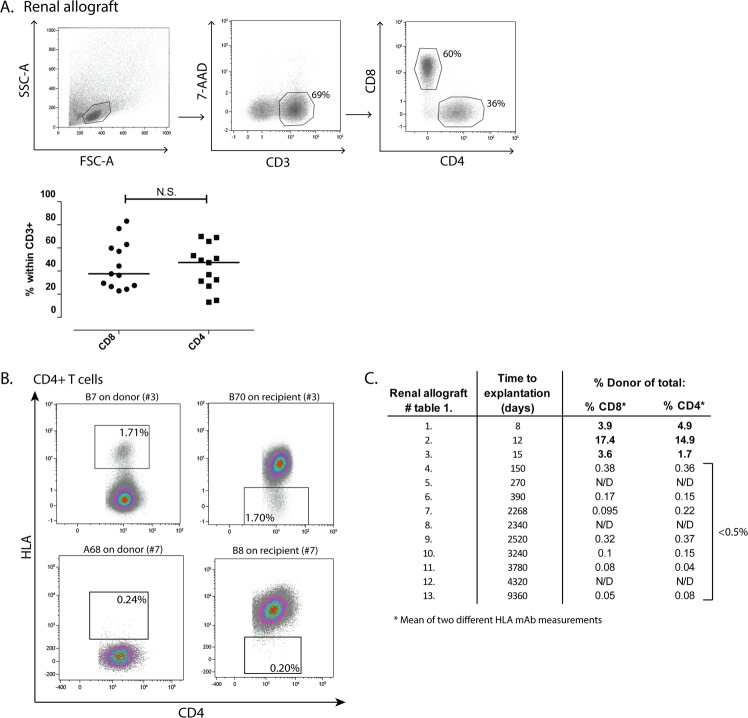
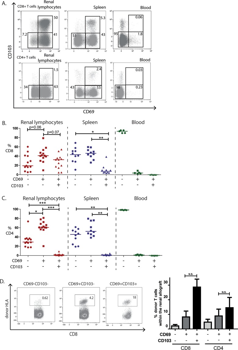
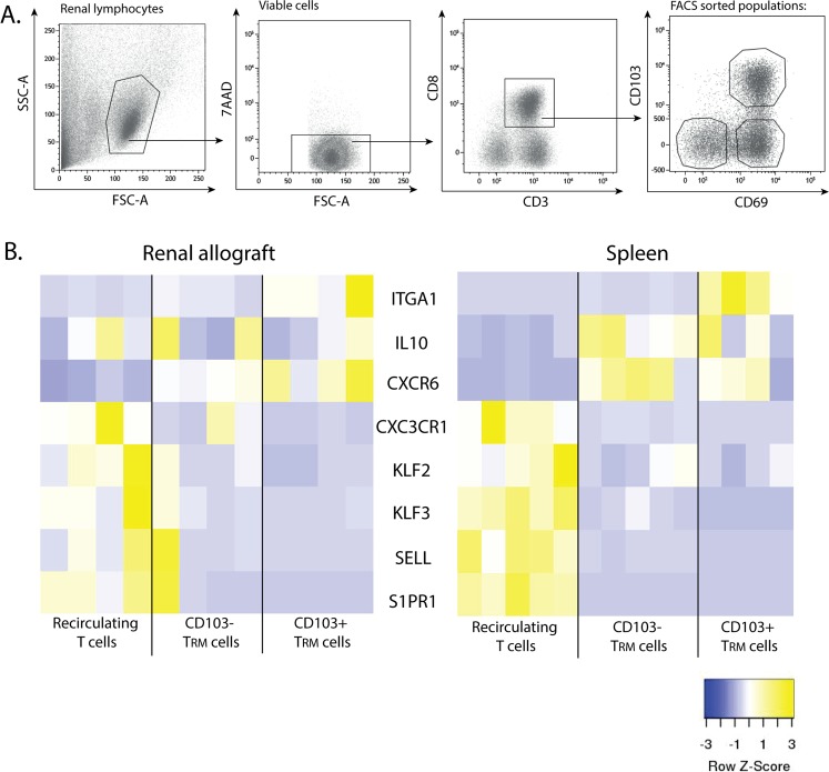
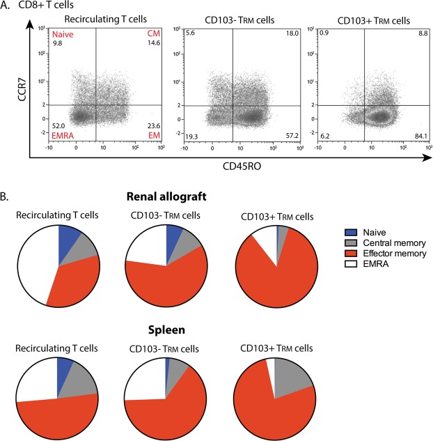
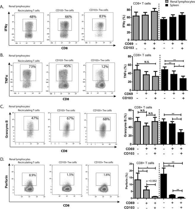
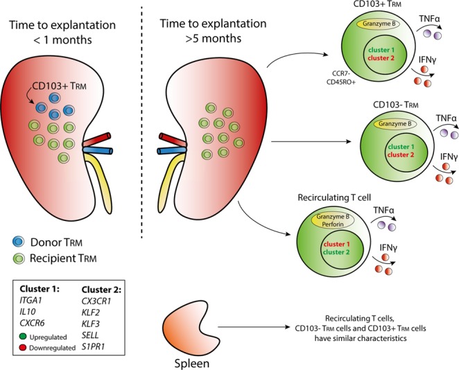
Tissue-resident memory T (TRM) cells are characterized by their surface expression of CD69 and can be subdivided in CD103+ and CD103- TRM cells. The origin and functional characteristics of TRM cells in the renal allograft are largely unknown. To determine these features we studied TRM cells in transplant nephrectomies. TRM cells with a CD103+ and CD103- phenotype were present in all samples (n = 13) and were mainly CD8+ T cells. Of note, donor-derived TRM cells were only detectable in renal allografts that failed in the first month after transplantation. Grafts, which failed later, mainly contained recipient derived TRM cells. The gene expression profiles of the recipient derived CD8+ TRM cells were studied in more detail and showed a previously described signature of tissue residence within both CD103+ and CD103- TRM cells. All CD8+ TRM cells had strong effector abilities through the production of IFNγ and TNFα, and harboured high levels of intracellular granzyme B and low levels of perforin. In conclusion, our results demonstrate that donor and recipient TRM cells reside in the rejected renal allograft. Over time, the donor-derived TRM cells are replaced by recipient TRM cells which have features that enables these cells to aggressively respond to the allograft.


Conflict of interest statement

D.A. Hesselink has received lecture and consulting fees from Astellas Pharma and Chiesi Farmaceutici SpA, as well as grant support from Astellas Pharma, Bristol-Myers Squibb, and Chiesi Farmaceutici SpA (paid to the Erasmus MC). F.J.M.F. Dor has received lecture and consulting fees from Astellas Pharma, Chiesi Farmaceutici SpA, Sandoz, and TEVA pharmaceuticals.

Figures

Figure 1
Figure 1
CD8+ and CD4+ T cells of donor and recipient origin are present in the renal allograft. Lymphocytes were isolated from the rejected renal allografts and subsequently stained and analysed by flow cytometry. (A) Gating strategy used to detect CD3+ T cells within the total viable lymphocytes, of which the CD8+ and CD4+ T cells were selected. (B) Typical examples of renal lymphocyte samples of patient number three and seven stained with mAb against human leukocyte antigen (HLA) class I antigens within the CD4+ T cells. Proportions of cells originating from the donor are depicted. (C) Table with numbers referring to the renal allografts described in Table 1, the time to explantation in days, and the proportions of donor cells detected within the CD8+ and CD4+ T cell compartment. Frequencies of the cells are presented as individual proportions with medians. HLA = human leukocyte antigen; N.S. = not significant; N/D = not determined.
Figure 2
Figure 2
TRM cells are present within CD8+ and CD4+ T cells in the renal allograft. Lymphocytes from rejected renal allografts, spleens of organ donors, and blood from healthy individuals were stained with mAb against CD69 and CD103. (A) Representative examples of the gating strategy of CD69 and CD103 of lymphocytes originating from the renal allograft, spleen, and blood. Proportions of the gated areas are depicted within the dot-plots. (B,C) Quantified data of the recirculating T cells (CD69−CD103−), CD103− TRM cells (CD69+CD103−), and CD103+ TRM cells (CD69+CD103+) subsets within the CD8+ T cell compartment (B) and CD4+ T cell compartment (C) of the renal allograft, spleen, and blood (blood n = 5, spleen n = 10, renal allograft n = 13). Frequencies of the cells are presented as individual proportions with medians. Significant differences were calculated and presented (*p < 0.05, **p < 0.01, ***p < 0.001). (D) mAb against HLA class I antigens were used to discriminate between donor and recipient lymphocytes. Typical example dot plots and quantified data of proportions of donor-derived cells within the CD103+ TRM cells, CD103− TRM cells and recirculating T cells are depicted of the transplant nephrectomies removed within the first month after transplantation (Table 1, patient 1 to 3). Frequencies of the positive cells are depicted within the dot plots. Frequencies of positive cells are shown as mean with the SEM (n = 3; N.S. = not significant).
Figure 3
Figure 3
Expression of TRM signature genes. (A) Typical example of the gating strategy used after fluorescence activated CD8+ cell sorting to obtain recirculating T cells (CD69−CD103−), CD103− TRM cells (CD69+CD103−), and CD103+ TRM cells (CD69+CD103+). Cells were gated by forward- and side-scatter followed by 7-AAD negative (viable) gating. (B) Heatmaps depicting the normalized gene expression of eight signature genes known to be upregulated (yellow) or downregulated (blue) in TRM cells of the renal allograft and spleen. Spleen n = 5, renal lymphocytes n = 4.
Figure 4
Figure 4
TRM cells in the renal allograft have an effector memory phenotype. (A) Typical examples of dot plots presenting the distribution of naïve T cells (CCR7+CD45RO−), central memory T cells (CM; CCR7+CD45RO+), effector memory T cells (EM; CCR7−CD45RO+), and EMRA T cells (CCR7−CD45RO−) within the CD8+ recirculating T cells, CD103− TRM cells and CD103+ TRM cells of the renal allograft. Numbers within the dot plots indicate proportions of the different cell subsets. (B) Pie charts representing the median proportion of naïve, central memory, effector memory, and EMRA T cells within the CD8+ recirculating T cells, CD103− TRM cells and CD103+ TRM cells of the renal allograft and spleen (renal lymphocytes n = 6, spleen n = 8).
Figure 5
Figure 5
TRM cells are capable of producing effector molecules. (A,B) Proportions of IFNγ (A) and TNFα (B) producing cells are depicted upon 4 hours PMA/ionomycin stimulation in the presence of monensin and brefeldin A. Cytokine proportions were measured in renal lymphocytes and splenocytes within the recirculating T cells, CD103− TRM cells, and CD103+ TRM cells of the CD8+ T cell compartment. (C,D) Frequencies of granzyme B (C) and perforin (D) levels were measured in the recirculating T cells, CD103− TRM, and CD103+ TRM cells of the CD8+ T cell compartment. Frequencies of positive cells were shown as mean with the SEM (renal lymphocytes n = 6, spleen n = 10). Significant differences were calculated and depicted (N.S. = not significant, *p < 0.05, **p < 0.01).
Figure 6
Figure 6
Schematic overview of TRM cell characteristics in the renal allograft. Distribution of donor-derived and recipient-derived tissue-resident memory (TRM) cells and the phenotypic and functional characteristics of the recipient-derived TRM cells in the explanted renal allograft are depicted in a schematic overview. Cluster one and cluster two indicate TRM core genes of which the expression was measured. Cluster one consists of genes involved in T cell activation (ITGA1, IL10, and CXCR6) and cluster two consists of genes involved in T cell migration (CX3CR1, KLF2, KLF3, SELL, S1PR1).

References

    1. Masopust D, Vezys V, Marzo AL, Lefrancois L. Preferential localization of effector memory cells in nonlymphoid tissue. Science. 2001;291:2413–2417. doi: 10.1126/science.1058867. - DOI - PubMed
    1. Park CO, Kupper TS. The emerging role of resident memory T cells in protective immunity and inflammatory disease. Nat Med. 2015;21:688–697. doi: 10.1038/nm.3883. - DOI - PMC - PubMed
    1. Woon HG, et al. Compartmentalization of Total and Virus-Specific Tissue-Resident Memory CD8+ T Cells in Human Lymphoid Organs. PLoS Pathog. 2016;12:e1005799. doi: 10.1371/journal.ppat.1005799. - DOI - PMC - PubMed
    1. Watanabe R, et al. Human skin is protected by four functionally and phenotypically discrete populations of resident and recirculating memory T cells. Sci Transl Med. 2015;7:279ra239. doi: 10.1126/scitranslmed.3010302. - DOI - PMC - PubMed
    1. Wakim LM, Woodward-Davis A, Bevan MJ. Memory T cells persisting within the brain after local infection show functional adaptations to their tissue of residence. Proc Natl Acad Sci USA. 2010;107:17872–17879. doi: 10.1073/pnas.1010201107. - DOI - PMC - PubMed

Publication types