

Fetal anogenital distance using ultrasound
- PMID: 30980419
- PMCID: PMC6618155
- DOI: 10.1002/pd.5459
Fetal anogenital distance using ultrasound
Abstract
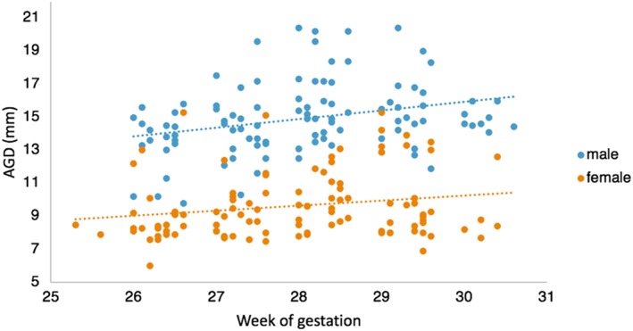
Objective: This study measured anogenital distance (AGD) during late second/early third trimester of pregnancy to confirm previous findings that AGD can be measured noninvasively in the fetus using ultrasound and further showed differences in reference ranges between populations.
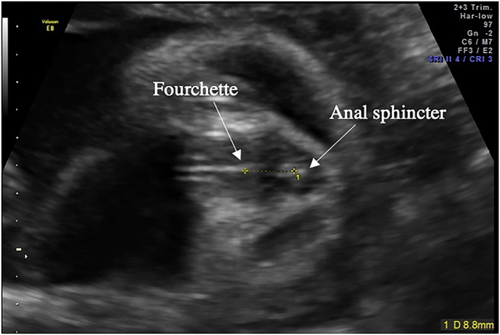
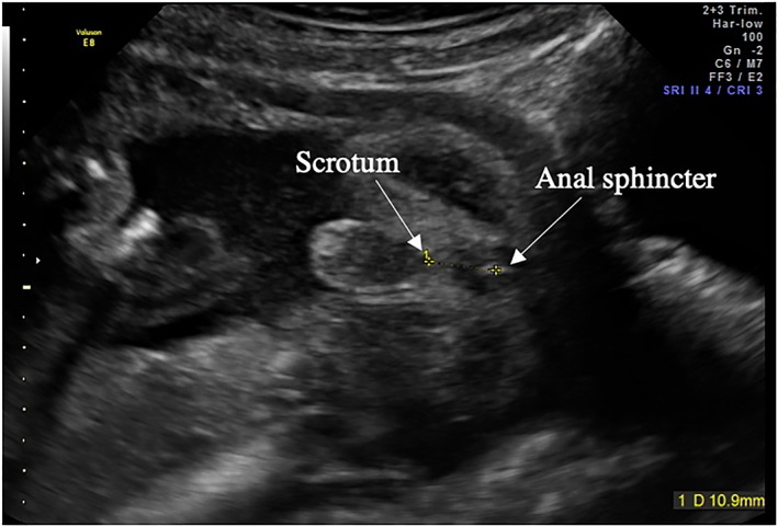
Method: Two hundred ten singleton pregnancies were recruited at the Rosie Hospital, Cambridge, UK. A 2D ultrasound was performed between 26 and 30 weeks of pregnancy. AGD was measured from the centre of the anus to the base of the scrotum in males and to the posterior convergence of the fourchette in females.
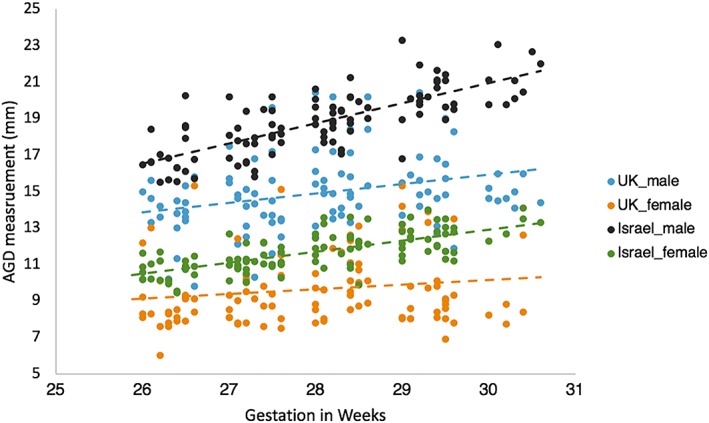
Results: A significant difference in AGD between males and females (P < .0001) was found, replicating previous results with a significant correlation between estimated fetal weight (EFW) and AGD in males only (P = .006). A comparison of AGD using reference data from an Israeli sample (n = 118) and our UK sample (n = 208) showed a significant difference (P < .0001) in both males and females, after controlling for gestational age (GA).
Conclusion: Our results confirm that AGD measurement in utero using ultrasound is feasible. In addition, there are strong sex differences, consistent with previous suggestions that AGD is influenced by prenatal androgen exposure. AGD lengths differ between the UK and Israel; therefore, population-specific normative values may be required for accurate clinical assessments.
© 2019 The Authors. Prenatal Diagnosis Published by John Wiley & Sons Ltd.
Conflict of interest statement
The authors have no potential conflicts of interest to disclose.
Figures
References
-
- Gilboa Y, Kivilevitch Z, Oren M, Cohen YP, Katorza E, Achiron R. Anogenital distance in male and female fetuses at 20 to 35weeks of gestation: centile charts and reference ranges. Prenat Diagn. 2014;34(10):946‐951. - PubMed
-
- Jain VG, Singal AK. Shorter anogenital distance correlates with undescended testis: a detailed genital anthropometric analysis in human newborns. Hum Reprod. 2013;28(9):2343‐2349. - PubMed
-
- Freire C, Ocón‐Hernández O, Dávila‐Arias C, et al. Anogenital distance and reproductive outcomes in 9‐ to 11‐year‐old boys: the INMA‐Granada cohort study. Andrology [Internet]. 2018;6(6):1‐8. Available from: http://doi.wiley.com/10.1111/andr.12544 - DOI - PubMed
MeSH terms
Grants and funding
LinkOut - more resources
Full Text Sources
