. 2019 Apr 13;16(1):86.
doi: 10.1186/s12974-019-1463-y.

Dysregulation of sonic hedgehog pathway and pericytes in the brain after lentiviral infection

Affiliations

Dysregulation of sonic hedgehog pathway and pericytes in the brain after lentiviral infection

Diana G Bohannon et al. J Neuroinflammation. .

Abstract

Background: Impairment of the blood-brain barrier (BBB) has been associated with cognitive decline in many CNS diseases, including HIV-associated neurocognitive disorders (HAND). Recent research suggests an important role for the Sonic hedgehog (Shh) signaling pathway in the maintenance of BBB integrity under both physiological and pathological conditions.

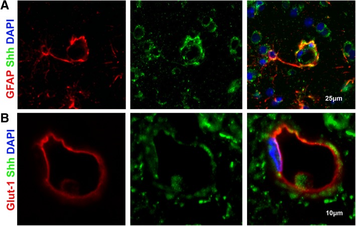
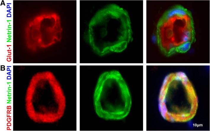
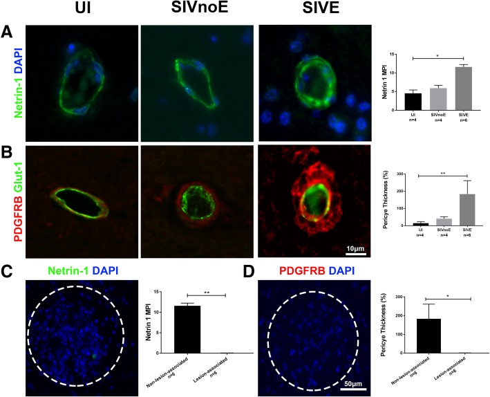
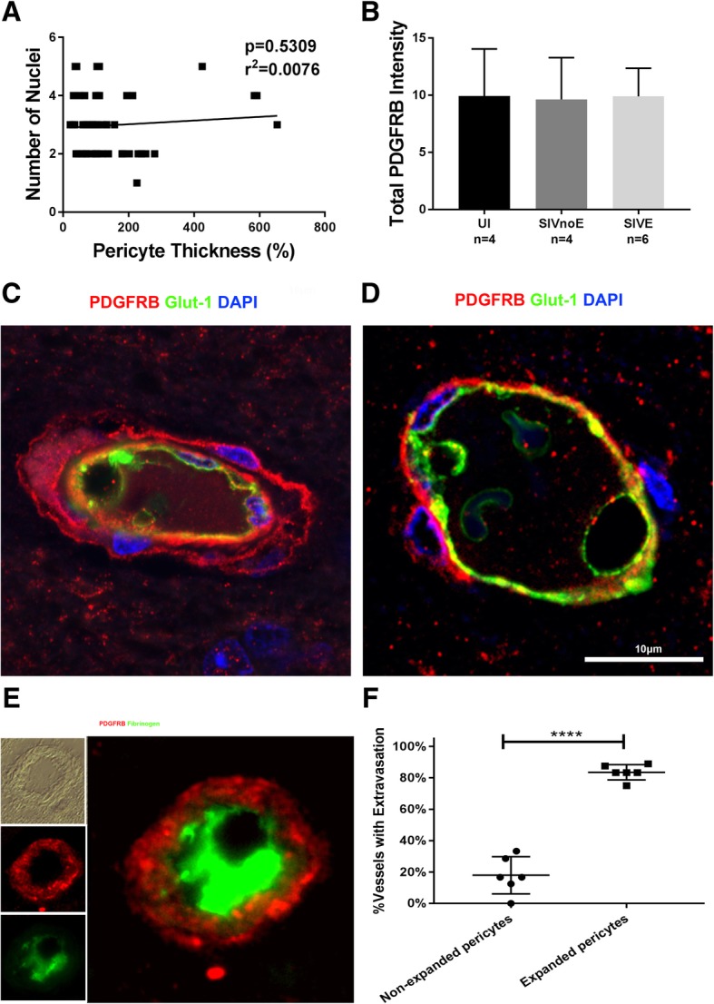
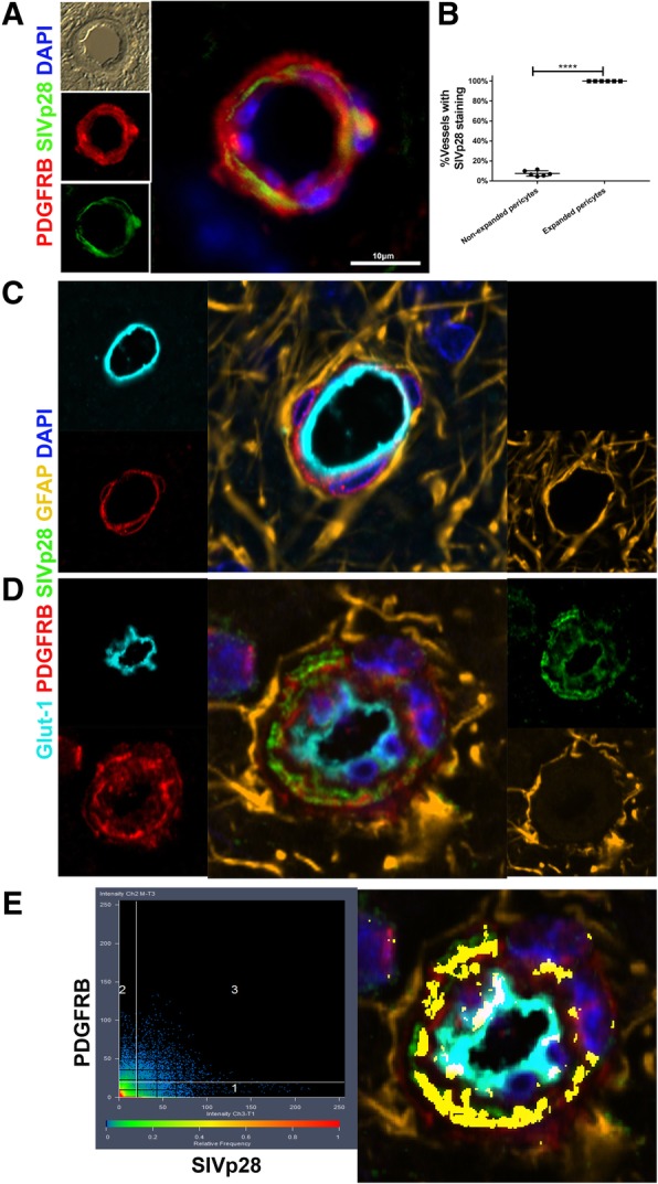
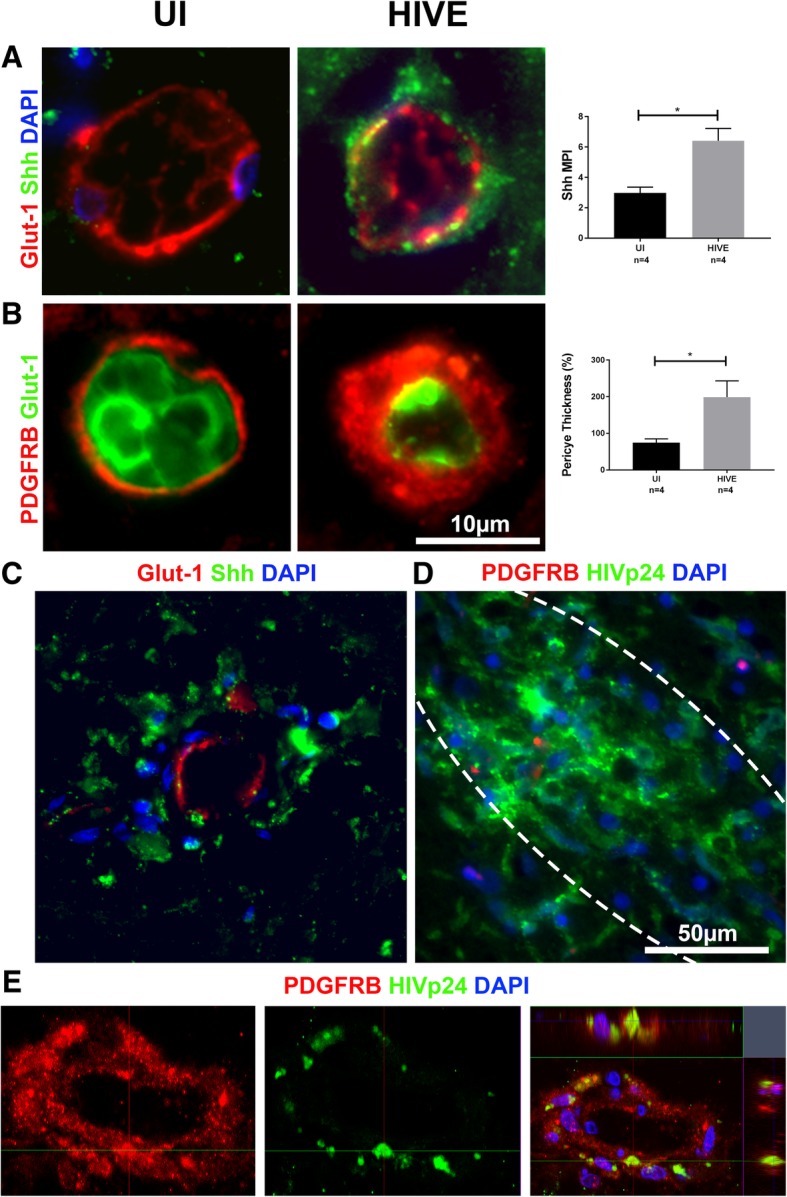
Methods: In the present study, we sought to examine the expression of Shh and its downstream effectors in relation to brain pericytes and BBB integrity in HIV-infected humans and rhesus macaques infected with simian immunodeficiency virus (SIV), an animal model of HIV infection and CNS disease. Cortical brain tissues from uninfected (n = 4) and SIV-infected macaques with (SIVE, n = 6) or without encephalitis (SIVnoE, n = 4) were examined using multi-label, semi-quantitative immunofluorescence microscopy of Shh, netrin-1, tight junction protein zona occludens 1 (ZO1), glial fibrillary acidic protein, CD163, platelet-derived growth factor receptor b (PDGFRB), glucose transporter 1, fibrinogen, and SIV Gag p28.

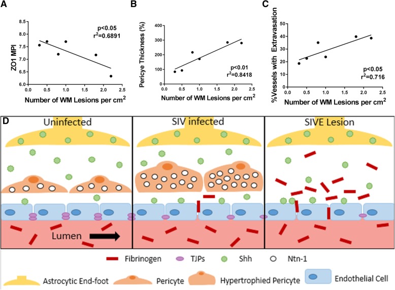
Results: While Shh presence in the brain persisted during HIV/SIV infection, both netrin-1 immunoreactivity and the size of PDGFRB+ pericytes, a cellular source of netrin-1, were increased around non-lesion-associated vessels in encephalitis compared to uninfected brain or brain without encephalitis, but were completely absent in encephalitic lesions. Hypertrophied pericytes were strongly localized in areas of fibrinogen extravasation and showed the presence of intracellular SIVp28 and HIVp24 by immunofluorescence in all SIV and HIV encephalitis cases examined, respectively.

Conclusions: The lack of pericytes and netrin-1 in encephalitic lesions, in line with downregulation of ZO1 on the fenestrated endothelium, suggests that pericyte loss, despite the strong presence of Shh, contributes to HIV/SIV-induced BBB disruption and neuropathogenesis in HAND.

Keywords: AIDS; Blood-brain barrier; HIV encephalitis; Netrin-1; Pericytes.


Conflict of interest statement

Consent for publication

Not applicable.

Competing interests

The authors declare that they have no competing interests.

Publisher’s Note

Springer Nature remains neutral with regard to jurisdictional claims in published maps and institutional affiliations.

Figures

Fig. 1
Fig. 1
Shh found on astrocytic end-feet and near endothelial cells within the glio-vascular unit triple-label IF imaging of uninfected animals for GFAP (red), Shh (green), and DAPI (blue) confirmed the presence of Shh on astrocytic end-feet (a). Likewise, IF imaging of uninfected animals for GLUT1 (red), Shh (green), and DAPI (blue) showed Shh located adjacent to endothelial cells b
Fig. 2
Fig. 2
Dysregulation of Shh with SIV infection double-label IF for Shh (green) and DAPI (blue) shows no significant difference between groups a. Triple-label IF of GLUT1 (red), Shh (green), and DAPI (blue) shows an increase in the MPI of Shh near the endothelium of both infection groups when compared to the control group (b). Likewise, IF imaging of Shh (green), GLUT1 (red), and DAPI (blue) in a SIVE lesion shows consistently high levels of Shh near the lesion-associated endothelium c. No significant difference was found in Shh levels between non-lesion-associated vessels and lesion-associated vessels in SIVE d. Twelve lesions were used from each SIVE animal (n = 6), and Shh MFI was compared between non-lesion and lesion-associated vessels in each animal. Lesions are circled with white dotted lines (c). Error bars denote SD
Fig. 3
Fig. 3
Netrin-1 found in pericytes within the neurovascular niche triple IF staining of uninfected animals for GLUT1 (red), netrin-1 (green), and DAPI (blue) shows netrin-1 surrounding the endothelium (a). Triple IF staining of uninfected animals for PDGFRB (red), netrin-1 (green), and DAPI (blue) shows netrin-1 co-localizing with pericytes at the glio-vascular interface b
Fig. 4
Fig. 4
Pericytes and netrin-1 dysregulated with SIV infection IF imaging of netrin-1 (green) and DAPI (blue) show an increase in netrin-1 MPI in SIVE animals (a). Double IF staining of PDGFRB (red) and GLUT1 (green) displays increased pericyte thickness in SIVE animals compared to uninfected (b). Double IF staining for PDGFRB (red), and DAPI (blue), shows that PDGFRB is eliminated in SIVE lesions (d). Likewise, staining for netrin-1 (green), and DAPI (blue), shows a similar elimination of netrin-1 in SIVE lesions c. Twelve lesions were used from each SIVE animal (n = 6) and PDGFRB area (c) or netrin-1 MPI (d) was compared between non-lesion and lesion-associated vessels in each animal. Lesions are circled within white dotted lines (c, d). Error bars denote SD
Fig. 5
Fig. 5
Changes in pericyte thickness of SIVE brain are due to cellular changes, not pericyte number. Pericyte thickness graphed against the number of pericytic nuclei shows no significant trend between the number of pericytes and their thickness around the endothelium of SIVE infected animals (a). Total PDGFRB intensity shows no change in PDGFRB expression between study groups despite the significant difference in pericyte area (b). Triple-label IF for PDGFRB (red), GLUT1 (green), and DAPI (blue) of an expanded pericyte (c) and a non-expanded pericyte (d) show the cellular differences between two pericytes in the same SIVE animal (10A067). Using double-label IF for PDGFRB (red) and fibrinogen (green), we were able to determine that 80% of vessels surrounded by enlarged pericytes showed signs of fibrinogen extravasation (e, f). Error bar denotes SD. Ten vessels were randomly chosen from each SIVE animal (n = 6); each point on the graph a denotes a single vessel. No significant difference was found in a or b. A paired t test was used to compare between samples in f
Fig. 6
Fig. 6
Enlarged pericytes highly localized to areas of fibrinogen extravasation and pericyte infection IF imaging for PDGFRB (red), SIVp28 (green), and DAPI (blue) a of 10 vessels with and 10 without enlarged pericyte coverage for each SIVE animal (n = 6) showed that 100% of the enlarged pericytes (n = 60) had localized SIVp28+ staining, while only 8% of the non-enlarged pericytes (n = 60) showed positive staining (b). Five-color IF staining for GFAP (gold), PDGFRB (red), GLUT1 (aqua), SIVp28 (green), and DAPI (dark blue) show the major cellular components of the glio-vascular interface and how they localize with SIVp28 viral protein marker in the non-hypertrophied pericyte of an uninfected animal (11A014) and the hypertrophied pericyte of an SIVE animal (11A554) (c, d). This demonstrates the presence of SIVp28 primarily in the pericytes and endothelial cells (d). Presence of SIVp28+ staining within pericytes was confirmed via a confocal microscopy Z-stack of a 20-μm-thick section using 0.25 μm increments analyzed with a ZenBlack co-localization test in which points in quadrant 3 and highlighted in yellow are considered to be co-localized between PDGFRB and SIVp28 (e). A paired t test was used to determine significance between groups in b
Fig. 7
Fig. 7
Findings in rhesus macaques confirmed in human tissue triple IF for Shh (green), GLUT1 (red), and DAPI (blue) show increased IR of Shh in association with the vessels of HIVE patients when compared to UI a and remained high in lesion-associated vessels (c). Likewise, double IF for GLUT1 (green) and PDGFRB (red) shows an increase in pericyte thickness in HIVE patients (b). Despite this increase, however, triple IF for HIVp24 (green), PDGFRB (red), and DAPI (blue) demonstrates a marked lack of PDGFRB+ pericytes in lesions (d). Further investigation of the same IF stain shows HIV Gag p24 localized to hypertrophied pericytes. The presence of HIVp24 within human brain pericytes was confirmed via a confocal microscopy Z-stack using 0.25 μm increments and an orthogonal view representation e. Lesions are circled with white dotted lines (d). A paired t test was used to determine significance between groups in a and b
Fig. 8
Fig. 8
BBB instability, pericyte thickness, and ZO1 loss correlate with the severity of SIVE. The severity of encephalitis was approximated by counting the average number of lesions per square cm of white matter tissue (n = 15 slides per SIVE animal). Each point on the graph designates the mean value of one SIVE animal (a–c). ZO1 MPI decreased in accordance with the lesion number (p < 0.05, r2 = 0.689) (a). The average pericyte area increases with lesion number (p < 0.01, r2 = 0.842) (b). The percent of vessels demonstrating fibrinogen extravasation increased with lesion number (p < 0.05, r2 = 0.716) (c). Lines demonstrate linear regressions (a–c). An illustrative representation of changes with the glio-vascular unit through disease progression depicts a section of the BBB in uninfected, SIV-infected, and SIVE animals (d). Uninfected animals have an intact endothelium with TJPs between endothelial cells, non-hypertrophied pericytes, fibrinogen confined within the lumen of brain vessels, Shh localized primarily in the open space pace between the endothelial cells and the astrocyte endfeet (d), left panel. SIV-infected animals show a partially fenestrated endothelium missing some TJPs, hypertrophied pericytes with increased netrin-1, some fibrinogen escaping the lumen, and Shh more heavily localized to the endothelium (d), middle panel. Within the lesion, encephalitic animals have a highly fenestrated endothelial layer with an almost complete loss of TJPs, high levels of fibrinogen extravasation, a complete loss of pericytes and netrin-1, and a high proportion of Shh localized to the endothelium (d), right panel

References

    1. Abbott NJ, Patabendige AA, Dolman DE, Yusof SR, Begley DJ. Structure and function of the blood-brain barrier. Neurobiol Dis. 2010;37:13–25. doi: 10.1016/j.nbd.2009.07.030. - DOI - PubMed
    1. Daneman R, Prat A. The blood-brain barrier. Cold Spring Harb Perspect Biol. 2015;7:a020412. doi: 10.1101/cshperspect.a020412. - DOI - PMC - PubMed
    1. Armulik A, Genove G, Mae M, Nisancioglu MH, Wallgard E, Niaudet C, et al. Pericytes regulate the blood-brain barrier. Nature. 2010;468:557–561. doi: 10.1038/nature09522. - DOI - PubMed
    1. Krueger M, Bechmann I. CNS pericytes: concepts, misconceptions, and a way out. Glia. 2010;58:1–10. doi: 10.1002/glia.20898. - DOI - PubMed
    1. Zhao Z, Nelson AR, Betsholtz C, Zlokovic BV. Establishment and dysfunction of the blood-brain barrier. Cell. 2015;163:1064–1078. doi: 10.1016/j.cell.2015.10.067. - DOI - PMC - PubMed

MeSH terms