Review
. 2019 Aug;138(2):221-236.
doi: 10.1007/s00401-019-02004-0. Epub 2019 Apr 13.

The new genetic landscape of Alzheimer's disease: from amyloid cascade to genetically driven synaptic failure hypothesis?

Affiliations
Review

The new genetic landscape of Alzheimer's disease: from amyloid cascade to genetically driven synaptic failure hypothesis?

Pierre Dourlen et al. Acta Neuropathol. 2019 Aug.

Abstract

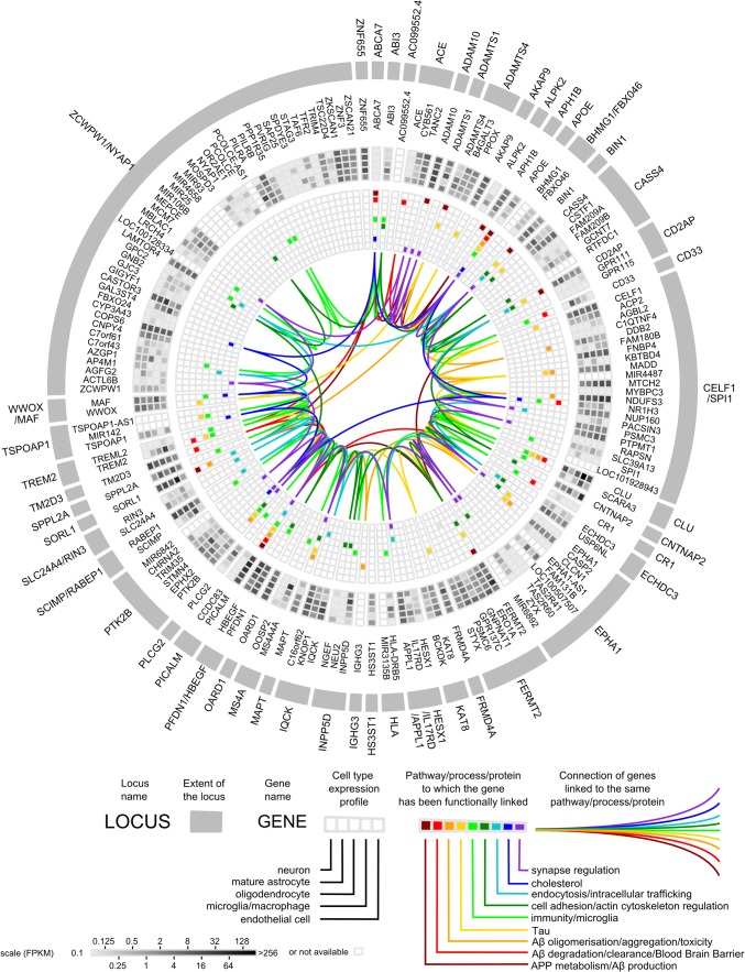
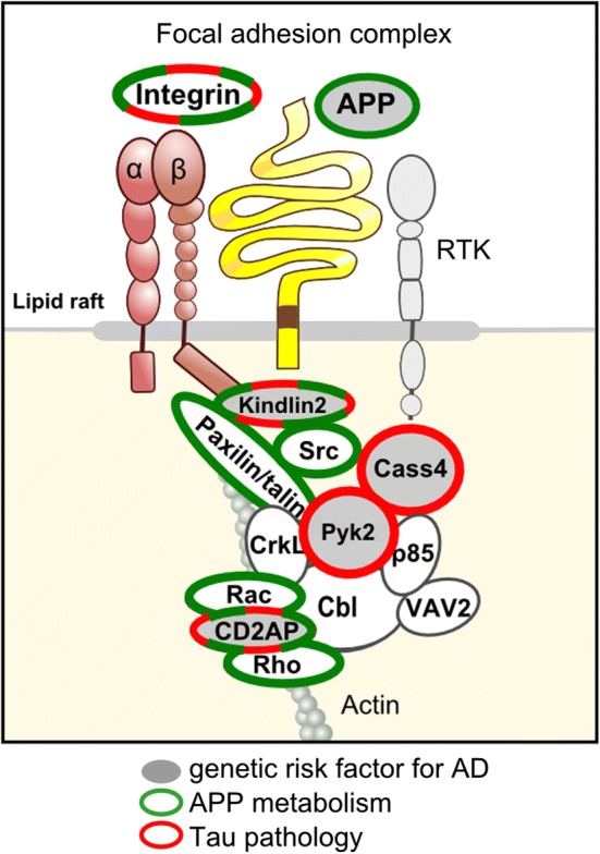
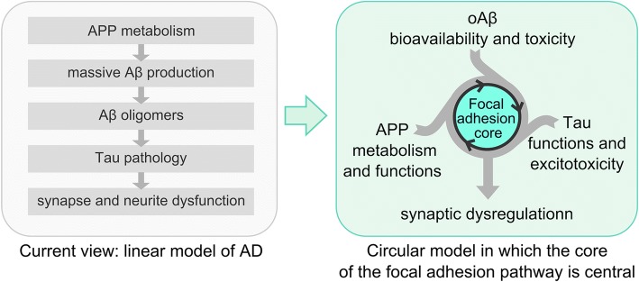
A strong genetic predisposition (60-80% of attributable risk) is present in Alzheimer's disease (AD). In view of this major genetic component, identification of the genetic risk factors has been a major objective in the AD field with the ultimate aim to better understand the pathological processes. In this review, we present how the genetic risk factors are involved in APP metabolism, β-amyloid peptide production, degradation, aggregation and toxicity, innate immunity, and Tau toxicity. In addition, on the basis of the new genetic landscape, resulting from the recent high-throughput genomic approaches and emerging neurobiological information, we propose an over-arching model in which the focal adhesion pathway and the related cell signalling are key elements in AD pathogenesis. The core of the focal adhesion pathway links the physiological functions of amyloid precursor protein and Tau with the pathophysiological processes they are involved in. This model includes several entry points, fitting with the different origins for the disease, and supports the notion that dysregulation of synaptic plasticity is a central node in AD. Notably, our interpretation of the latest data from genome wide association studies complements other hypotheses already developed in the AD field, i.e., amyloid cascade, cellular phase or propagation hypotheses. Genetically driven synaptic failure hypothesis will need to be further tested experimentally within the general AD framework.


Figures

Fig. 1
Fig. 1
Circular diagram of AD genetic risk factors. The diagram shows (from outside to inside): (1) genomic loci in alphabetical order; (2) genes therein; (3) expression profiles of these genes in different cell types of the brain (grayscale); and (4) the pathways/processes/proteins to which these genes have been functionally linked (colour). Details of the functional studies supporting these linkages are available in Suppl. Table. Expression profiles were extracted from [155] (FPKM fragments per kilobase of transcript sequence per million mapped fragments). The circlize package of the R software (http://www.r-project.org/) was used to generate the diagram [38]
Fig. 2
Fig. 2
Amyloid cascade hypothesis and genetic risk factors of AD. Autosomal dominant mutations that cause early onset familial AD (in APP, PSEN1 and PSEN2) gave rise to the amyloid cascade hypothesis, which aims to link amyloid plaques and neurofibrillary tangles, the two classical AD hallmarks. Involvement of the genetic risk factors of late-onset AD in APP metabolism and in Aβ clearance through the blood brain barrier or microglia supports this hypothesis. Soluble forms of Aβ may be inducing neurotoxicity through modifying Tau metabolism, leading to neurofibrillary tangle formation and neuronal death. Recent GWAS-defined genes that modulate Tau toxicity may be involved in Aβ-induced neurotoxicity through mechanisms that are yet to be identified
Fig. 3
Fig. 3
Interactions between integrin, APP and receptor tyrosine kinases (RTKs) at the cell surface modulate cell adhesion. The GWAS-defined genes FERMT2, Cass4, PTK2B and CD2AP, recently identified for their roles in APP metabolism (in green) and/or Tau pathology (in red), are involved in the focal adhesion complex, which regulates several downstream cell signalling pathways as well as the actin cytoskeleton. This observation supports the concept that the focal adhesion core, together with its related downstream pathways, may be an important actor in the AD process
Fig. 4
Fig. 4
Genetic risk factors and synapse dysfunction in AD pathogenesis. Regulation of the focal adhesion pathway plays central roles in synaptic plasticity (synaptic maintenance, actin cytoskeleton remodelling, vesicle, and receptor cycling). Dysfunction of downstream cellular signalling pathways involving APP and/or Tau may thus participate in synapse loss. Additionally, dysregulation of the core FA pathway could modulate APP and Tau metabolisms, leading to an exacerbation of synaptotoxicity through Aβ overproduction and Tau-modulated excitotoxicity. Finally, Aβ availability at the synapses is dependent on its clearance through the blood brain barrier and/or by microglial cells
Fig. 5
Fig. 5
Reappraisal of the amyloid cascade hypothesis into a circular model. In this new model, dysfunction of the FA pathway at synapses could be the basis of a vicious cycle with multiple entry points linking AD hallmarks to synapse dysregulation, synapse loss, and subsequent cognitive decline

References

    1. Andersen OM, Reiche J, Schmidt V, Gotthardt M, Spoelgen R, Behlke J, et al. Neuronal sorting protein-related receptor sorLA/LR11 regulates processing of the amyloid precursor protein. Proc Natl Acad Sci USA. 2005;102:13461–13466. doi: 10.1073/pnas.0503689102. - DOI - PMC - PubMed
    1. Andrew RJ, De Rossi P, Nguyen P, Kowalski HR, Recupero AJ, Guerbette T, et al. Reduction of the expression of the late-onset Alzheimer’s disease (AD) risk-factor BIN1 does not affect amyloid pathology in an AD mouse model. J Biol Chem. 2019;294:4477–4487. doi: 10.1074/jbc.ra118.006379. - DOI - PMC - PubMed
    1. Atagi Y, Liu C-C, Painter MM, Chen X-F, Verbeeck C, Zheng H, et al. Apolipoprotein E is a ligand for triggering receptor expressed on myeloid cells 2 (TREM2) J Biol Chem. 2015;290:26043–26050. doi: 10.1074/jbc.m115.679043. - DOI - PMC - PubMed
    1. Bailey CC, Devaux LB, Farzan M. The triggering receptor expressed on myeloid cells 2 binds apolipoprotein E. J Biol Chem. 2015;290:26033–26042. doi: 10.1074/jbc.m115.677286. - DOI - PMC - PubMed
    1. Bales KR, Verina T, Cummins DJ, Du Y, Dodel RC, Saura J, et al. Apolipoprotein E is essential for amyloid deposition in the APP(V717F) transgenic mouse model of Alzheimer’s disease. Proc Natl Acad Sci USA. 1999;96:15233–15238. doi: 10.1073/pnas.96.26.15233. - DOI - PMC - PubMed

Publication types

MeSH terms