Review
. 2019 Nov 15;13(6):617-627.
doi: 10.5009/gnl18476.

Characterization of Intraductal Papillary Neoplasm of the Bile Duct with Respect to the Histopathologic Similarities to Pancreatic Intraductal Papillary Mucinous Neoplasm

Affiliations
Review

Characterization of Intraductal Papillary Neoplasm of the Bile Duct with Respect to the Histopathologic Similarities to Pancreatic Intraductal Papillary Mucinous Neoplasm

Yasuni Nakanuma et al. Gut Liver. .

Abstract

Intraductal papillary neoplasms of the bile duct (IPNBs) are known to show various pathologic features and biological behaviors. Recently, two categories of IPNBs have been proposed based on their histologic similarities to pancreatic intraductal papillary mucinous neoplasms (IPMNs): type 1 IPNBs, which share many features with IPMNs; and type 2 IPNBs, which are variably different from IPMNs. The four IPNB subtypes were re-evaluated with respect to these two categories. Intestinal IPNBs showing a predominantly villous growth may correspond to type 1, while those showing papillay-tubular or papillay-villous growth correspond to type 2. Regarding gastric IPNB, those with regular foveolar structures with varying numbers of pyloric glands may correspond to type 1, while those with papillary-foveolar structures with gastric immunophenotypes and complicated structures may correspond to type 2. Pancreatobiliary IPNBs that show fine ramifying branching may be categorized as type 1, while others containing many complicated structures may be categorized as type 2. Oncocytic type, which displays solid growth or irregular papillary structures, may correspond to type 2, while papillary configurations with pseudostratified oncocytic lining cells correspond to type 1. Generally, type 1 IPNBs of any subtype develop in the intrahepatic bile ducts, while type 2 IPNBs develop in the extrahepatic bile duct. These findings suggest that IPNBs arising in the intrahepatic ducts are biliary counterparts of IPMNs, while those arising in the extrahepatic ducts display differences from prototypical IPMNs. The recognition of these two categories of IPNBs with reference to IPMNs and their anatomical location along the biliary tree may deepen our understanding of IPNBs.

Keywords: Biliary anatomy; Cholangiocarcinoma; Counterpart; Intraductal papillary mucinous neoplasm of pancreas; Intraductal papillary neoplasm of bile duct.


Conflict of interest statement

CONFLICTS OF INTEREST

No potential conflict of interest relevant to this article was reported.

Figures

Fig. 1
Fig. 1
Intraductal papillary neoplasm of the bile duct arising in the extrahepatic bile duct. (A) A papillary lesion and laterally spreading lesion in the extrahepatic bile duct (H&E, ×50). (B) A higher-magnification view of the papillary lesions marked by circle in A shows fine fibrovascular cores covered by neoplastic epithelia (H&E, ×70). (C) A higher-magnification view of the laterally spreading lesion marked by circle in A shows micropapillary and flat neoplastic epithelia (H&E, ×100).
Fig. 2
Fig. 2
The gross features of an intraductal papillary neoplasm of the bile duct (IPNB). (A) An IPNB growing in the intrahepatic bile duct showing cystic dilatation (white asterisk, dilated bile duct lumen). (B) IPNB in the extrahepatic bile duct showing villous features (one tumor was separated into two [arrows] at the time of dissection).
Fig. 3
Fig. 3
Intestinal-type intraductal papillary neoplasm of the bile duct (IPNB) (type 1). (A) An IPNB arising in the intrahepatic bile duct shows a villous architecture with thin fibrovascular cores (H&E, ×80). (B) A higher-magnification view of the villous neoplasm shows stratified nuclei with amphiphilic cytoplasm and little supranuclear mucin (H&E, ×120).
Fig. 4
Fig. 4
Intestinal-type intraductal papillary neoplasm of the bile duct (IPNB) (type 2). (A) An IPNB arising in the distal bile duct shows papillary-tubular architecture with thin, focally fibrotic, fibrovascular cores (H&E, ×60). (B) An IPNB arising in the distal bile duct with high-grade dysplasia. The structures of the papillary-tubular patterns are irregular (H&E, ×80). (C) These neoplastic cells are diffusely positive for CDX2 (immunostaining, ×70).
Fig. 5
Fig. 5
Gastric-type intraductal papillary neoplasm of the intrahepatic bile duct (type 1). Foveolar epithelia and pyloric glands comprise the basic architecture of this gastric neoplasm (H&E, ×150).
Fig. 6
Fig. 6
Gastric-type intraductal papillary neoplasm of the extrahepatic bile duct (type 2). (A) A papillary neoplasm with fine fibrovascular cores and epithelial lining (asterisk), and flat and nonpapillary neoplastic epithelial lining in the adjacent mucosa (arrow) are identifiable (H&E, ×80). (B) A higher-magnification view of (A) shows a low columnar clear cytoplasm and basally located nuclei appearing as pyloric glands (right half) and low columnar epithelia with less mucinous cytoplasm, reminiscent of gastric foveola (left half). Asterisk indicates solid structures (H&E, ×120).
Fig. 7
Fig. 7
Pancreatobiliary-type intraductal papillary neoplasm of the bile duct (IPNB) (type 1). (A) An IPNB arising in the intrahepatic bile duct shows fine and branching fibrovascular cores lined by columnar epithelia. (H&E, ×120). (B) Fine and branching stalks are lined by low columnar and cuboidal epithelia. A higher-magnification view of (A) (H&E, ×140).
Fig. 8
Fig. 8
Pancreatobiliary-type intraductal papillary neoplasm of the bile duct (IPNB) (type 2). (A) An IPNB arising in the extrahepatic bile duct showing fine and branching fibrovascular stalks lined by papillary neoplastic epithelia (H&E, ×50). (B) The epithelial lining shows branching stalks with papillary, pseudopapillary and serrated epithelia with high-grade dysplasia (H&E, ×120).
Fig. 9
Fig. 9
Oncocytic-type intraductal papillary neoplasm of the intrahepatic bile duct (type 1). (A) A papillary neoplasm with fine fibrovascular cores and epithelial lining (H&E, ×50). (B) Lining the epithelia are several cell layers with numerous secondary lumina and acidophilic or oncocytic cytoplasm as well as hyperchromatic nuclei. A higher-magnification view of (A) (H&E, ×120).
Fig. 10
Fig. 10
Oncocytic-type intraductal papillary neoplasm of the extrahepatic bile duct (type 2). (A) A papillary neoplasm shows polypoid growth with fine fibrovascular cores and multilayered nuclei (H&E, ×40). (B) The papillary or compact growth of oncocytic cells with hyperchromatic and pleomorphic nuclei. The secondary lumina are vague (asterisks, edematous stalk). A higher-magnification view of (A) (H&E, ×150).
Fig. 11
Fig. 11
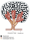
The development of the intrahepatic bile duct, extrahepatic bile duct and hepatocytes differs in mice (18.5 embryonic days). The hepatocytes are directly derived from albumin-positive hepatoblasts in the hepatic diverticulum, while the intrahepatic bile ducts are derived via the ductal plate from albumin-positive hepatoblasts in the hepatic diverticulum, and the extrahepatic bile duct is derived from albumin-negative hepatoblasts in the hepatic diverticulum. Adapted with permission from Nishikawa Y and Enomoto K.

References

    1. Nakanuma Y, Curado M-P, Franceschi S, et al. Intrahepatic cholangiocarcinoma. In: Bosman FT, Carneiro F, Hruban RH, Theise ND, editors. WHO of tumours of the digestive system. Lyon: International Agency for Research on Cancer; 2010. pp. 217–224.
    1. Albores-Saavedra J, Adsay NV, Crawford JM, et al. Carcinoma of the gallbladder and extrahepatic bile ducts. In: Bosman FT, Carneiro F, Hruban RH, Theise ND, editors. WHO classification of tumours of the digestive system. Lyon: International Agency for Research on Cancer; 2010. pp. 266–273.
    1. Chen TC, Nakanuma Y, Zen Y, et al. Intraductal papillary neoplasia of the liver associated with hepatolithiasis. Hepatology. 2001;34(4 Pt 1):651–658. doi: 10.1053/jhep.2001.28199. - DOI - PubMed
    1. Albores-Saavedra J, Murakata L, Krueger JE, Henson DE. Noninvasive and minimally invasive papillary carcinomas of the extrahepatic bile ducts. Cancer. 2000;89:508–515. doi: 10.1002/1097-0142(20000801)89:3<508::AID-CNCR5>3.0.CO;2-D. - DOI - PubMed
    1. Nakanuma Y, Kakuda Y, Uesaka K, et al. Characterization of intraductal papillary neoplasm of bile duct with respect to histopathologic similarities to pancreatic intraductal papillary mucinous neoplasm. Hum Pathol. 2016;51:103–113. doi: 10.1016/j.humpath.2015.12.022. - DOI - PubMed

MeSH terms