. 2019 May 2;104(5):936-947.
doi: 10.1016/j.ajhg.2019.03.010. Epub 2019 Apr 11.

Homozygous Mutations in CSF1R Cause a Pediatric-Onset Leukoencephalopathy and Can Result in Congenital Absence of Microglia

Affiliations

Homozygous Mutations in CSF1R Cause a Pediatric-Onset Leukoencephalopathy and Can Result in Congenital Absence of Microglia

Nynke Oosterhof et al. Am J Hum Genet. .

Abstract

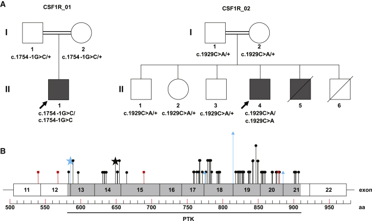
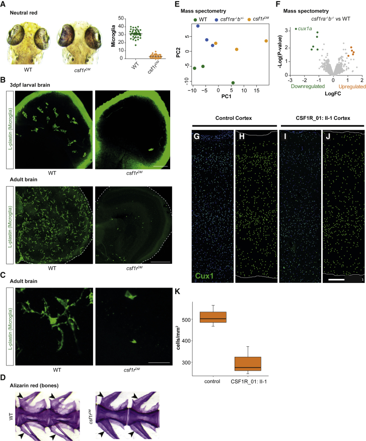
Microglia are CNS-resident macrophages that scavenge debris and regulate immune responses. Proliferation and development of macrophages, including microglia, requires Colony Stimulating Factor 1 Receptor (CSF1R), a gene previously associated with a dominant adult-onset neurological condition (adult-onset leukoencephalopathy with axonal spheroids and pigmented glia). Here, we report two unrelated individuals with homozygous CSF1R mutations whose presentation was distinct from ALSP. Post-mortem examination of an individual with a homozygous splice mutation (c.1754-1G>C) demonstrated several structural brain anomalies, including agenesis of corpus callosum. Immunostaining demonstrated almost complete absence of microglia within this brain, suggesting that it developed in the absence of microglia. The second individual had a homozygous missense mutation (c.1929C>A [p.His643Gln]) and presented with developmental delay and epilepsy in childhood. We analyzed a zebrafish model (csf1rDM) lacking Csf1r function and found that their brains also lacked microglia and had reduced levels of CUX1, a neuronal transcription factor. CUX1+ neurons were also reduced in sections of homozygous CSF1R mutant human brain, identifying an evolutionarily conserved role for CSF1R signaling in production or maintenance of CUX1+ neurons. Since a large fraction of CUX1+ neurons project callosal axons, we speculate that microglia deficiency may contribute to agenesis of the corpus callosum via reduction in CUX1+ neurons. Our results suggest that CSF1R is required for human brain development and establish the csf1rDM fish as a model for microgliopathies. In addition, our results exemplify an under-recognized form of phenotypic expansion, in which genes associated with well-recognized, dominant conditions produce different phenotypes when biallelically mutated.

Keywords: CSF1R; CUX1; agenesis corpus callosum; axonal spheroids; leukoencephalopathy; microglia; neuropathology; osteopetrosis; recessive; zebrafish.


Figures

Figure 1
Figure 1
Pedigrees and Distribution of CSF1R Mutations (A) Pedigrees of family CSF1R_01 and CSF1R_02 shown, with genotypes indicated. CSF1R sequencing was not performed for II-5, but this individual is shaded to reflect his leukodystrophy and periventricular calcifications. (B) Exons 11-22 of the CSF1R protein (GenBank: NP_005202.2) are shown. No mutations have been reported in the five immunoglobin (Ig) domains and transmembrane domain upstream of exon 11, which are not shown. The intracellular protein tyrosine kinase (PTK) domain is indicated. The 64 previously reported pathogenic variants causative for ALSP are shown (truncating variants in red squares, missense and in-frame indel variants in black circles, splice variants in blue triangles). The splice site variant identified in family CSF1R_01 (GenBank: NM_005211.3; c.1754−1G>C) is indicated by the blue star. The missense variant identified in family CSF1R_02 (GenBank: NM_005211.3; c.1929C>A [p.His643Gln]) is indicated by the black star. Amino acids containing multiple variants are specified by proportionally taller lines. A complete list of reported CSF1R variants is in Table S1.
Figure 2
Figure 2
Brain and Bone Abnormalities in CSF1R Deficiency Shown are MRI images of individual II-1 (at age 2 days) from family CSF1R_01 (A–F) and MRI images of individual II-4 (at age 25 years) from family CSF1R_02 (G–L). (A)–(E) are T1 weighted images; (F)–(H) are T2 weighted; (I) and (K) are T1 FLAIR images; and (J) and (L) are T2 FLAIR images. (A) Midsagittal section shows agenesis of corpus callosum (asterisk), pontocerebellar hypoplasia (arrowhead), and small and upwardly rotated cerebellar vermis (arrow) with large posterior fossa cyst (“X”). (B) Parasagittal section shows diffuse calcifications along lateral ventricles (arrowhead) and small cerebellum and posterior fossa cyst (arrow). Note calcifications were confirmed on CT imaging (not shown) and gross pathology (Figure S4). (C) Horizontal section shows periventricular calcification around 3rd and lateral ventricles (arrow and asterisks, respectively). (D) Coronal section showing ventriculomegaly (asterisks), hypoplasia of cerebellar hemispheres and large posterior fossa cyst (arrow). (E and F) Periventricular white matter signal abnormality, with areas of T2 hyperintensity (arrow in F) with corresponding T1 hypointensity (arrow in E). (G) Midsagittal section showing cerebellar vermis hypoplasia/atrophy and hypoplasia of corpus callosum (arrow). (H) Parasagittal section showing periventricular T2 hyperintensities (arrow). (I–L) Axial T1 FLAIR (I and K) and corresponding T2 FLAIR images (J and L) showing frontal lobe predominant periventricular white matter signal abnormality, with areas of T2/FLAIR hyperintensity with corresponding T1 hypointensity (arrows). (M–O) X-rays of individual II-1 from family CSF1R_01 showing generalized increased bone density and metaphyseal dysplasia (arrows).
Figure 3
Figure 3
Microscopic Brain Abnormalities in CSF1R Deficiency Sections from individual II-1 from family CSF1R_01 (A–E, I–K, O–Q); sections from control brain (F–H, L–N). (A and B) Adjacent sections through right frontal cortex, stained with H&E (A) and neurofilament protein immunohistochemistry (B). The white matter contained a focus of calcification, necrosis, and axon loss (arrowheads). (C) Deep cerebellar white matter contained disorganized heterotopia (arrowheads) beneath the cerebellar cortex, and multiple periventricular foci of calcification and severe gliosis (arrows). Stained with H&E. (D and E) Neurofilament immunohistochemistry demonstrated axonal spheroids in right frontal white matter. Similar axonal spheroids seen in anterior and lateral corticospinal tracts and nucleus cuneatus (not shown). (F–H) Iba1 immunohistochemistry in control white matter labeled numerous microglia with long, ramified processes insinuating through brain tissue. (G) and (H) are enlarged 2-fold relative to (F). (I–K) Iba1 immunohistochemistry in white matter from individual II-1 revealed decreased numbers of microglia, with abnormal rounded morphology, located mainly in perivascular spaces. (J) and (K) are enlarged 2-fold relative to (I). (L–N) CD68 immunohistochemistry in control white matter. (M) and (N) are enlarged 2-fold relative to (L). (O–Q) CD68 immunohistochemistry in white matter from individual II-1. Black arrows indicate blood vessels with no CD68-positive cells; the red arrow (O, enlarged in Q) indicates a rare perivascular CD68-positive cell. (P) and (Q) are enlarged 2-fold relative to (O). Scale bars: (A) 1 cm for (A) and (B); (C) 1 cm for (C) only; (D) 20 μm for (D)–(E); 40 μm for (F), (L), (I), (O); 20 μm for (G), (H), (J), (K), (M), (N), (P), (Q).
Figure 4
Figure 4
Analysis of csf1rDM Zebrafish and CSF1R-Deficient Human Cortex (A) Neutral red staining and quantification of microglia in zebrafish larvae (5 days after fertilization). (B) Microglia immunostaining (L-plastin) in larval brains and adult brain sections of control and csf1rDM zebrafish. (C) Microglia morphology in adult control and csf1rDM zebrafish (L-plastin immunostaining). (D) Alizarin red staining of vertebrae and arches of adult control and csf1rDM zebrafish (size-matched, age 6–8 months) Neural arch (upper arrow) and hemal arch (lower arrow) indicated (E). Principal component analysis (PCA) of mass spectrometry data of WT, csf1ra−/−; b+/−, and csf1rDM adult zebrafish brains. Csf1ra−/−; b+/− also have reduced microglia numbers. (F) Volcano plot showing differentially regulated proteins in csf1rDM mutant brains as colored dots (FDR < 0.05, LogFC > |1|). Volcano plot of csf1ra−/−; b+/− versus control brain is included as Figure S2. (G–K) CUX1+ cells were reduced in the cerebral cortex of CSF1R_01. CUX1 immunofluorescence (green) in control (G, H) and CSF1R-deficient (I, J) lateral temporal neocortex. The images with DAPI (blue) counterstain are shown in (G) and (I) and corresponding CUX1+ cell plots in (H) and (J). (K) The density of CUX1+ cells was reduced in the cortex from three different areas of individual II-1, family CSF1R_01. Cortical regions in this analysis included left medial parietal, left lateral parietal, and left lateral temporal cortex (control) and left lateral temporal, right dorsolateral frontal, and right medial occipital cortex (CSF1R_01). Whiskers indicate 95% confidence interval, and difference between CSF1R_01:II-1 and control CUX1+ cell density was statistically significant (p < 0.05, one-sided t test, and p = 0.05, one-sided exact Wilcoxon rank sums test).

References

    1. Cunningham C.L., Martínez-Cerdeño V., Noctor S.C. Microglia regulate the number of neural precursor cells in the developing cerebral cortex. J. Neurosci. 2013;33:4216–4233. - PMC - PubMed
    1. Erblich B., Zhu L., Etgen A.M., Dobrenis K., Pollard J.W. Absence of colony stimulation factor-1 receptor results in loss of microglia, disrupted brain development and olfactory deficits. PLoS ONE. 2011;6:e26317. - PMC - PubMed
    1. Hagemeyer N., Hanft K.M., Akriditou M.A., Unger N., Park E.S., Stanley E.R., Staszewski O., Dimou L., Prinz M. Microglia contribute to normal myelinogenesis and to oligodendrocyte progenitor maintenance during adulthood. Acta Neuropathol. 2017;134:441–458. - PMC - PubMed
    1. Paolicelli R.C., Bolasco G., Pagani F., Maggi L., Scianni M., Panzanelli P., Giustetto M., Ferreira T.A., Guiducci E., Dumas L. Synaptic pruning by microglia is necessary for normal brain development. Science. 2011;333:1456–1458. - PubMed
    1. Squarzoni P., Oller G., Hoeffel G., Pont-Lezica L., Rostaing P., Low D., Bessis A., Ginhoux F., Garel S. Microglia modulate wiring of the embryonic forebrain. Cell Rep. 2014;8:1271–1279. - PubMed

Publication types

MeSH terms

Substances