Review
. 2019 Jun;72(6):325-349.
doi: 10.1038/s41429-019-0171-2. Epub 2019 Apr 15.

Targeting adenylate-forming enzymes with designed sulfonyladenosine inhibitors

Affiliations
Review

Targeting adenylate-forming enzymes with designed sulfonyladenosine inhibitors

Michaelyn C Lux et al. J Antibiot (Tokyo). 2019 Jun.

Abstract

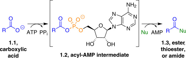
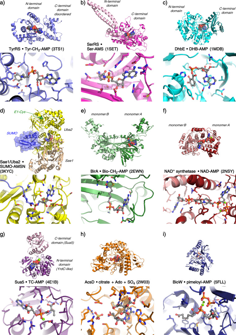
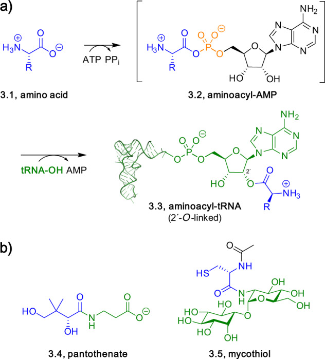
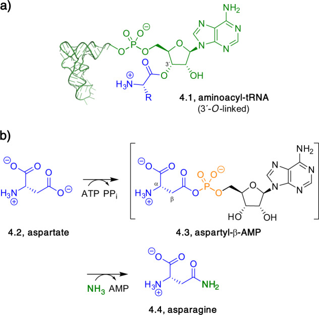
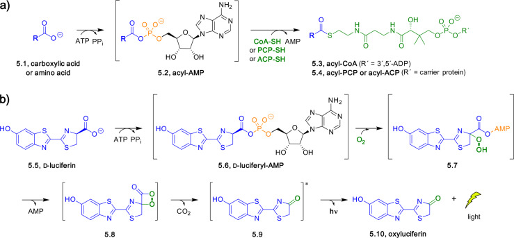
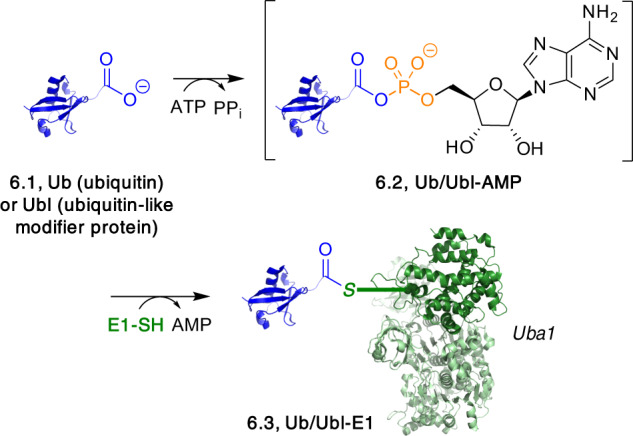
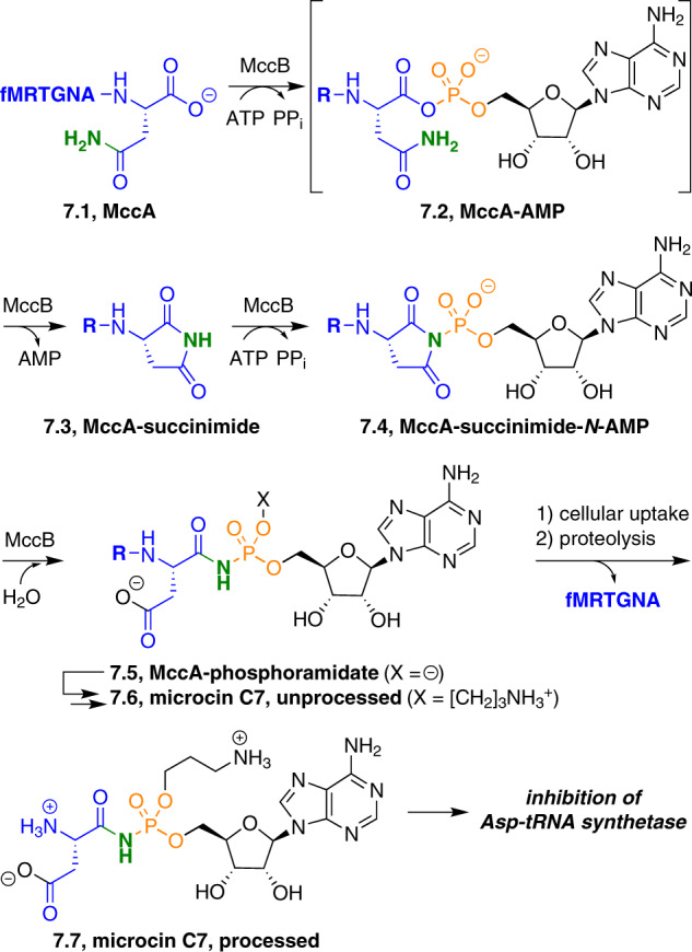
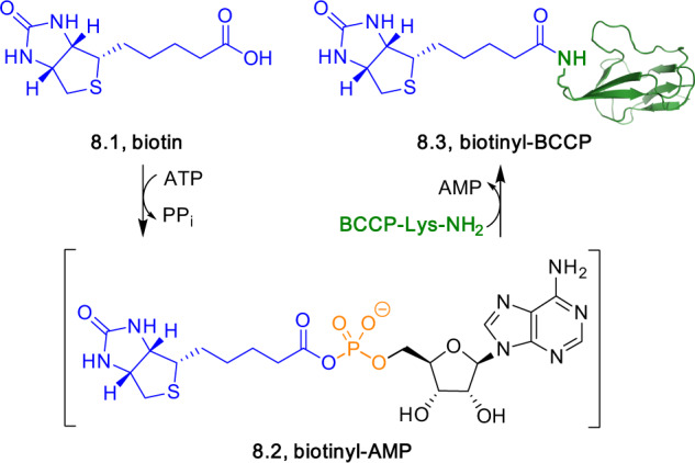
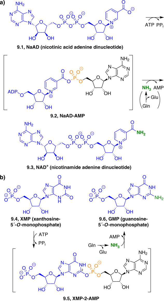
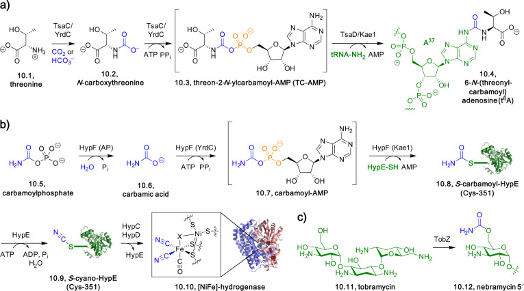
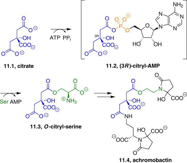
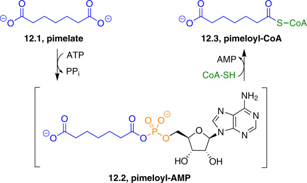
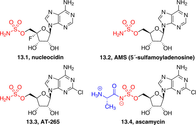
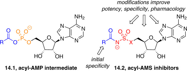
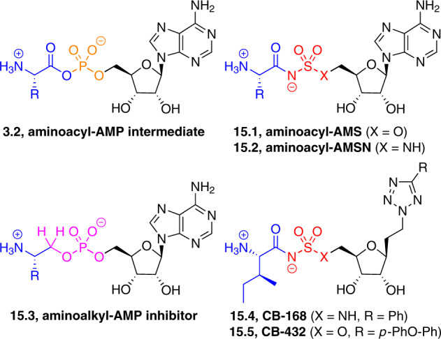
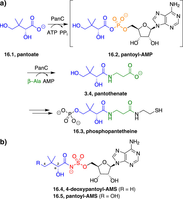
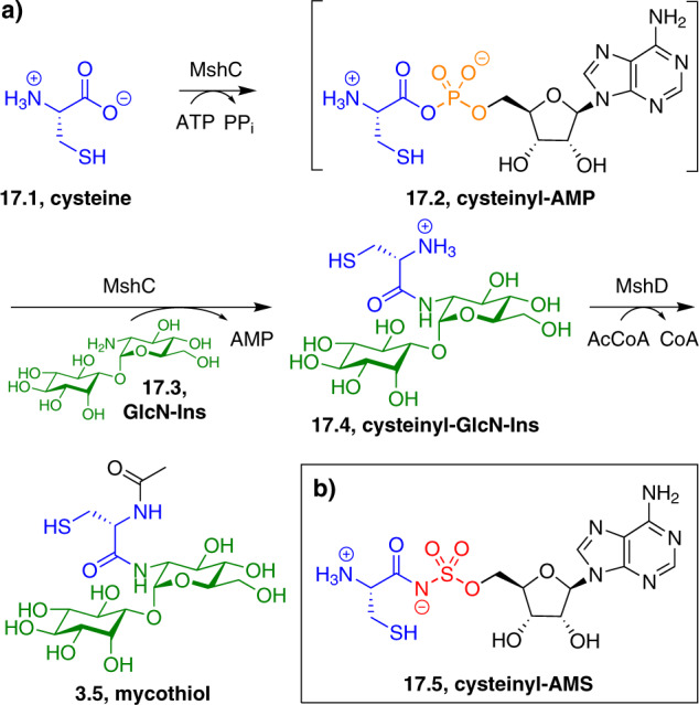
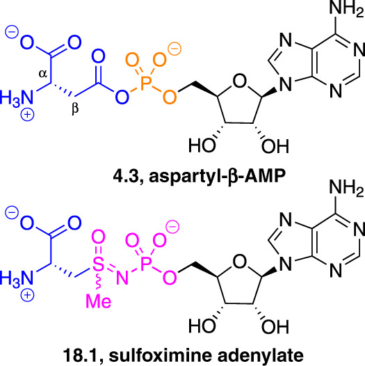
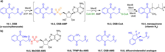
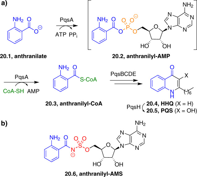
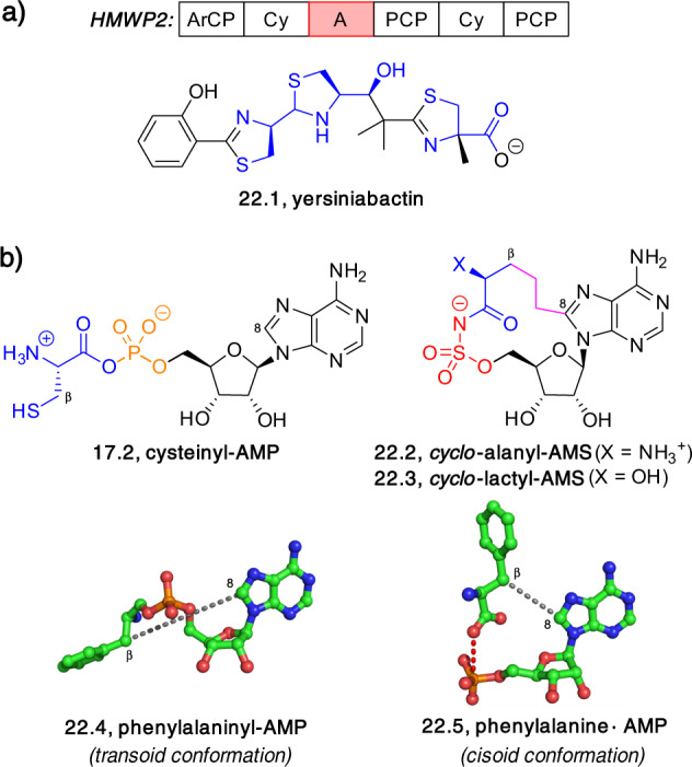
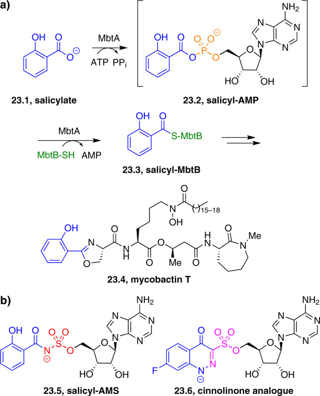
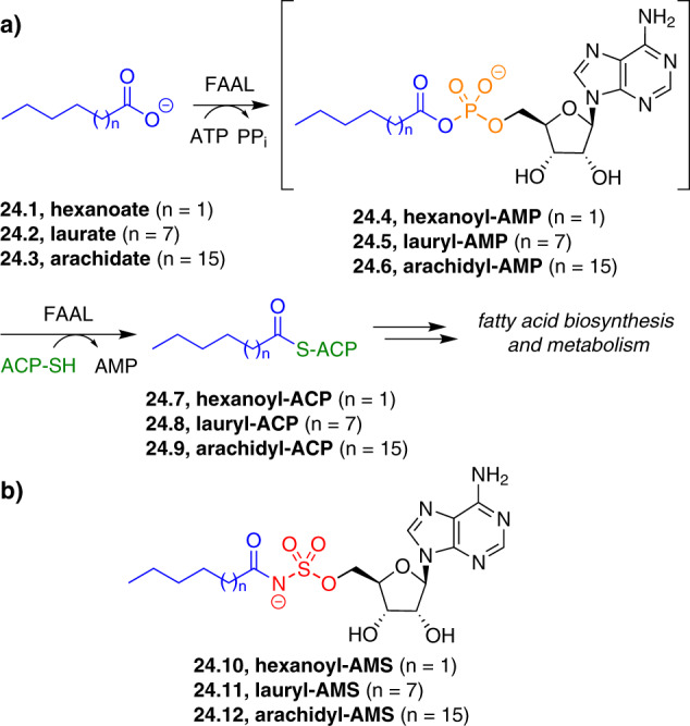
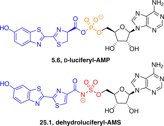
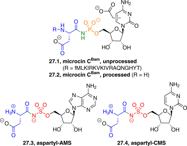
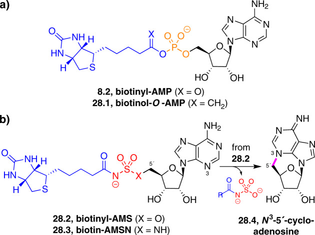
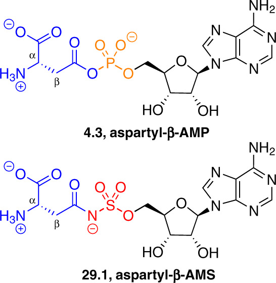
Adenylate-forming enzymes are a mechanistic superfamily that are involved in diverse biochemical pathways. They catalyze ATP-dependent activation of carboxylic acid substrates as reactive acyl adenylate (acyl-AMP) intermediates and subsequent coupling to various nucleophiles to generate ester, thioester, and amide products. Inspired by natural products, acyl sulfonyladenosines (acyl-AMS) that mimic the tightly bound acyl-AMP reaction intermediates have been developed as potent inhibitors of adenylate-forming enzymes. This simple yet powerful inhibitor design platform has provided a wide range of biological probes as well as several therapeutic lead compounds. Herein, we provide an overview of the nine structural classes of adenylate-forming enzymes and examples of acyl-AMS inhibitors that have been developed for each.


Conflict of interest statement

D.S.T. is a coinventor of U.S. Patents 8,461,128 and 8,946,188; International Patent Applications PCT/US2016/055136 and PCT/US2016/055200; and U.S. Provisional Patent Applications 62/527,925, 62/527,932, 62/527,936, 62/527,943, 62/784,323, and 62/802,650 concerning sulfonyladenosine analogues. L.C.S. is a coinventor of U.S. Provisional Patent Application 62/784,323. M.C.L. declares no conflicts of interest.

Figures

Fig. 1
Fig. 1
General mechanism for two half-reactions catalyzed by adenylate-forming enzymes. A carboxylic acid substrate (1.1) (blue) attacks ATP at the α-phosphate (orange) to form a reactive acyl-AMP (acyl adenylate) intermediate (1.2), which then reacts with a nucleophile (green) to form an ester, thioester, or amide product (1.3). In some cases, the second half-reaction is accompanied by a conformational change in the enzyme that leads to active-site remodeling. AMP adenosine-5′-O-monophosphate, ATP adenosine-5′-O-triphosphate
Fig. 2
Fig. 2
Structures of the nine classes of adenylate-forming enzymes. Top of ah: Overall protein fold with acyl-AMP intermediate or mimic (spheres) bound. Bottom of ah: Active site with acyl-AMP intermediate or mimic (ball-and-stick) bound; protein side chains within 4 Å of the ligand are shown (sticks); portions of structures that obscure the ligands are not shown. PDB ID codes are shown in parentheses. a Class I aminoacyl-tRNA synthetase Bacillus stearothermophilus tyrosyl-tRNA synthetase catalytic N-terminal domain with carbonyl-reduced intermediate mimic Tyr-CH2-AMP (tyrosinyl-AMP) (PDB ID: 3TS1) [44]. b Class II aminoacyl-tRNA synthetase Thermus thermophilus seryl-tRNA synthetase with adenylate mimic Ser-AMS (seryl-AMS) (PDB ID: 1SET) [134]. c ANL family enzyme B. subtilis DhbE with adenylate intermediate DHB-AMP (2,3-dihydroxybenzoyl-AMP) (PDB ID: 1MDB) [55]. d SUMO (small ubiquitin-like modifier) E1 activating enzyme human (Sae1/Uba2) with adenylate mimic SUMO-AMSN (SUMO1[T95C]-AMSN) (PDB ID: 3KYC) [60]. e Biotin protein ligase Escherichia coli BirA homodimer with carbonyl-reduced intermediate mimic Bio-CH2-AMP (biotinol-O-AMP) (PDB ID: 2EWN) [66]. f N-type ATP pyrophosphatase B. subtilis NAD+ synthetase homodimer with adenylate intermediate NAD-AMP (PDB ID: 2NSY) [68]. g YrdC-like carbamoyltransferase Sulfolobus tokodaii Sua5 with adenylate intermediate TC-AMP (threon-2-N-ylcarbamoyl-AMP) (PDB ID: 4E1B) [79, 80]. h NRPS-independent siderophore synthetase Petrobacterium chrysanthemi AcsD with substrate citrate and ATP fragments Ado (adenosine) and SO4 (sulfate) (PDB ID: 2W03) [91]. i BioW pimeloyl-CoA synthetase with adenylate intermediate pimeloyl-AMP (PDB ID: 5FLL) [40]. Abbreviations: AMP adenosine-5′-O-monophosphate, AMS adeonsine-5′-O-monosulfamate, AMSN 5′-(aminodeoxy)adenosine-5′-N-monosulfamide, ANL acyl-CoA synthetase/NRPS adenylation domain/luciferase, ATP adenosine-5′-O-triphosphate, CoA coenzyme A, NAD nicotinamide adenine dinucleotide, NRPS non-ribosomal peptide synthetase, tRNA transfer ribonucleic acid
Fig. 3
Fig. 3
Biochemistry of class I aminoacyl-tRNA synthetases and related enzymes. a An amino acid (3.1) is adenylated to form an aminoacyl-AMP intermediate (3.2), which reacts with a tRNA 3′-ribose hydroxyl nucleophile (typically the 2′-hydroxyl) to form an aminoacyl-tRNA ester (3.3). b Pantothenate (3.4) and mycothiol (3.5) biosyntheses involve adenylate-forming enzymes having the same fold as class I aminoacyl-tRNA synthetases. Carboxylic acid substrate-derived fragments are shown in blue; nucleophile-derived fragments are shown in green. tRNA structure derived from PDB ID: 1GTR [213]
Fig. 4
Fig. 4
Biochemistry of class II aminoacyl-tRNA synthetases and related enzymes. a Aminoacyl-tRNA products (4.1) are produced by the same mechanism as in class I aminoacyl-tRNA synthetases (Fig. 3a), but are typically linked to the 3′-hydroxyl nucleophile of the tRNA 3′-ribose. b Asparagine synthetase A catalyzes adenylation of aspartate (4.2) at its β-carboxylate to form an aspartyl-β-AMP intermediate (4.3), which reacts with an ammonia nucleophile to form the primary carboxamide in asparagine (4.4). tRNA structure derived from PDB ID: 1ASY [214]
Fig. 5
Fig. 5
Biochemistry of ANL family enzymes. a Acyl-CoA synthetases and NRPS adenylation domains catalyze adenylation of carboxylic acids and amino acids (5.1) to form acyl-AMP intermediates (5.2), which react with a thiol nucleophile on the phosphopantetheine moiety of CoA or a carrier protein domain, respectively, to form thioester products (5.3, 5.4). b Firefly luciferase catalyzes adenylation of d-luciferin (5.5) to form a luciferyl-AMP intermediate (5.6), which reacts via the corresponding enolate with molecular oxygen to form an α-peroxide intermediate (5.7). Cyclization of the intramolecular peroxide nucleophile forms a peroxylactone (5.8). Fragmentation forms excited oxyluciferin (5.9), which emits light and returns to the ground state (5.10). ACP acyl carrier protein, PCP peptidyl carrier protein
Fig. 6
Fig. 6
Biochemistry of ubiquitin-family E1 activating enzymes. The C-terminal carboxylate of Ub or a Ubl (6.1) is adenylated to form a Ub/Ubl-AMP intermediate (6.2), which reacts with a remote catalytic cysteine side-chain thiol nucleophile on the E1 enzyme to form a Ub/Ubl-E1 thioester conjugate (6.3). Ub and Ub E1 (Uba1) structures derived from PDB ID: 4NNJ [215]. Ub ubiquitin, Ubl ubiquitin-like modifier protein
Fig. 7
Fig. 7
Biochemistry of E. coli microcin C7 synthetase MccB. The C-terminal carboxylate of the MccA precursor peptide (7.1) is adenylated to form a MccA-AMP intermediate (7.2), whose asparagine side-chain amide cyclizes to form a succinimide intermediate (7.3). MccB then catalyzes a second adenylation reaction to form a succinimide adenylate (7.4), which is hydrolyzed to form a phosphoramidate product (7.5). Downstream installation of an O-aminopropyl group provides microcin C (7.6). This Trojan horse antibiotic is taken up by target cells via peptide transporters, then the N-terminal peptide is proteolyzed to form an aspartyl-phosphoramidate (7.7), which inhibits aspartyl-tRNA synthetases in the target cell. R = fMRTGNA = N-formyl-Met-Arg-Thr-Gly-Asn-Ala peptide
Fig. 8
Fig. 8
Biochemistry of biotin protein ligases. Biotin (8.1) is adenylated to form a biotinyl-AMP intermediate (8.2), which reacts with a lysine side-chain amine nucleophile on the BCCP subunit of acetyl-CoA carboxylase to form an amide product (8.3). In the case of the E. coli enzyme BirA, formation of the adenylate intermediate (8.2) also induces homodimerization of the enzyme, which binds to and represses the bioO biosynthetic operon for biotin. BCCP structure derived from PDB ID: 1BDO [216]. BCCP biotin carboxylate carrier protein
Fig. 9
Fig. 9
Biochemistry of N-type ATP pyrophosphatases. a In a canonical mechanism using a carboxylic acid substrate, NAD+ synthetase catalyzes adenylation of NaAD (9.1) to form a NaAD-AMP intermediate (9.2), which reacts with an ammonia nucleophile to form a NAD+ primary carboxamide product (9.3). The ammonia is typically, but not always, derived from hydrolysis of the side-chain amide of glutamine (Gln → Glu + NH3) by a separate glutamine amidotransferase domain or subunit. b In a mechanistic variant using a non-carboxylic acid substrate, GMP synthetase catalyzes adenylation of the urea oxygen of XMP (9.4) to form an XMP-2-AMP O-adenylyl isourea intermediate (9.5), which reacts with an ammonia nucleophile to form a GMP guanidine product (9.6). GMP guanosine-5′-O-monophosphate, NaAD nicotinic acid adenine dinucleotide, XMP xanthosine 5′-O-monophosphate
Fig. 10
Fig. 10
Biochemistry of YrdC-like carbamoyltransferases. a In t6A biosynthesis, TsaC/YrdC converts threonine (10.1) to N-carboxythreonine (10.2), then adenylates it to form a TC-AMP intermediate (10.3). A separate enzyme, TsaD/Kae1, condenses the adenylate with the 6-amino group of tRNA-A37 to form t6A (10.4). b In [NiFe]-hydrogenase maturation, the multidomain protein HypF carries out AP-domain-catalyzed hydrolysis of carbamoylphosphate (10.5) to carbamic acid (10.6), YrdC-like domain-catalyzed adenylation to form a carbamoyl-AMP intermediate (10.7) and Kae1-like domain-catalyzed condensation with the C-terminal Cys-351 side-chain thiol nucleophile of HypE to form an S-thiocarbamate intermediate (10.8). Downstream enzymes then catalyze dehydration to a thiocyanate intermediate (10.9), cyanide transfer to an iron center, and installation of the complex in the [NiFe]-hydrogenase active site (10.10, X = bridging ligand). c The natural product tobramycin (10.11) is O-carbamoylated by TobZ, which contains YrdC-like and Kae1-like domains, to form nebramycin 5′ (10.12). HypE structure derived from PDB ID: 3VTI [84]; [NiFe]-hydrogenase structure derived from PDB ID: 2FRV [217]. AP acyl phosphatase domain, t6A 6-N-(threon-2-N-ylcarbamoyl)adenosine, TC threon-2-N-ylcarbamoyl
Fig. 11
Fig. 11
Biochemistry of NRPS-independent siderophore synthetases. This family typically uses diacid or monofunctionalized derivatives of diacids as substrates and couples them to alcohol or amine nucleophiles. P. crysanthemi AcsD catalyzes desymmetrizing adenylation of citrate (11.1) to form a (3R)-citryl-AMP intermediate (11.2), which reacts with a serine side-chain hydroxyl nucleophile to form an ester product (11.3), en route to the siderophore achromobactin (11.4)
Fig. 12
Fig. 12
Biochemistry of BioW acyl-CoA synthetases. Pimelate (12.1) is adenylated to form a pimeloyl-AMP intermediate (12.2), which reacts with a CoA thiol nucleophile to form a pimeloyl-CoA thioester product (12.3), en route to biotin
Fig. 13
Fig. 13
Sulfamoyladenosine (AMS) natural products
Fig. 14
Fig. 14
General acyl-AMS inhibitor design platform. Acyl-AMP intermediates (14.1) are generally bound tightly by the corresponding adenylate-forming enzymes and can be mimicked with non-hydrolyzable acyl-AMS analogues (14.2, X = O or NR). Initial specificity for the desired adenylate-forming enzyme is provided by the acyl group (blue) and additional modifications can be made in the acyl, sulfamate (red), ribose, and adenine regions in analogues
Fig. 15
Fig. 15
Inhibition of Class I aminoacyl-tRNA synthetases. Structures of aminoacyl-AMP reaction intermediates (3.2) and related adenylate-mimetic inhibitors (15.115.5)
Fig. 16
Fig. 16
Inhibition of panthothenate synthetase (PanC). a PanC catalyzes adenylation of pantoate (16.1) to form a pantoyl-AMP intermediate (16.2), which reacts with a β-alanine amine nucleophile to form a pantothenate amide product (3.4), en route to phosphopantetheine (16.3). b A 4-deoxypantoyl-AMS inhibitor (16.4) mimics the pantoyl-AMP intermediate (16.2) but lacks the 4-hydroxy group of pantoyl-AMS (16.5) to avoid decomposition via lactonization of the pantoyl side chain. *Stereochemistry epimeric or as shown
Fig. 17
Fig. 17
Inhibition of mycobacterial cysteine ligase (MshC). a MshC catalyzes adenylation of cysteine (17.1) to form a cysteinyl-AMP intermediate (17.2), which reacts with a GlcN-Ins amine nucleophile (17.3) to form a cysteinyl-GlcN-Ins amide product (17.4), en route to mycothiol (3.5). b A cysteinyl-AMS inhibitor (17.5) mimics the cysteinyl-AMP intermediate (17.2). GlcN-Ins 1-d-myo-inosityl-2-amido-2-deoxy-α-d-glucopyranoside
Fig. 18
Fig. 18
Inhibition of asparagine synthetase A. Structures of aspartyl-β-AMP reaction intermediate (4.3) and transition-state mimetic sulfoximine adenylate inhibitor (18.1)
Fig. 19
Fig. 19
Inhibition of bacterial OSB-CoA synthetase (MenE). a MenE catalyzes adenylation of OSB (19.1) to form an OSB-AMP intermediate (19.2), which reacts with a CoA thiol nucleophile to form an OSB-CoA thioester product (19.3), en route to menaquinone (19.4). b Acyl-AMS inhibitors (19.5–19.8) mimic the OSB-AMP intermediate (19.2). Various modifications have been explored to replace the anionic aromatic carboxylate of OSB-AMS (19.7). OSB o-succinylbenzoate, TFMP-Bu trifluoromethylphenyl-4-oxobutyryl
Fig. 20
Fig. 20
Inhibition of P. aeruginosa anthranilyl-CoA synthetase (PqsA). a PqsA catalyzes adenylation of anthranilate (20.1) to form an anthranilyl-AMP intermediate (20.2), which reacts with a CoA thiol nucleophile to form an anthranilyl-CoA thioester product (20.3), en route to the quinolone quorum-sensing factors HHQ (20.4) and PQS (20.5). b An anthranilyl-AMS inhibitor (20.6) mimics the anthranilyl-AMP intermediate (20.2). HHQ 2-heptyl-4-hydroxy-quinoline, PQS Pseudomonas quinolone signal = 3,4-dihydroxy-2-heptylquinoline
Fig. 21
Fig. 21
Inhibition of adenylation domains from NRPS pathways. a The GrsA subunit PheA domain catalyzes adenylation of l-phenylalanine (21.1) to form an l-phenylalanyl-AMP intermediate (21.2), which reacts with the GrsA PCP domain phosphopantetheine thiol nucleophile to form an acyl-PCP thioester product (21.3). The GrsA Ep domain then inverts the α-stereocenter of the thioester to form the d-congener (21.4). Peptide extension by the GrsB subunit and dimerization forms gramicidin S (21.5). Inset: The SrfA-C subunit LeuA domain catalyzes an analogous adenylation of l-leucine en route to surfactin (21.6). b Aminoacyl-AMS inhibitors mimic the aminoacyl-AMP intermediates of GrsA PheA (21.7) and SrfA-C LeuA (21.8), respectively. Ep epimerase, GrsA gramicidin synthetase A, LeuA leucine adenylation, PheA phenylalanine adenylation, SrfA-C surfactin synthetase C, TE thioesterase
Fig. 22
Fig. 22
Inhibition of yersiniabactin synthethase subunit HMWP2. a The yersiniabactin synthethase HMWP2 subunit cysteine A domain catalyzes adenylation of l-cysteine en route to yersiniabactin (22.1), where it is incorporated three times (blue). b Both HMWP2 and cysteinyl-tRNA synthetase form the same cysteinyl-AMP reaction intermediate (17.2), but aminoacyl-tRNA synthetases bind their adenylate intermediates in extended or transoid conformations (22.4) (phenylalaninyl-AMP bound to phenylalanyl-tRNA synthetase, from PDB ID: 1B7Y) [218], whereas NRPS adenylation domains bind them in cisoid conformations (22.5) (phenylalanine and AMP bound to GrsA PheA, from PDB ID: 1AMU) [54]. The macrocyclic constraint (red) in cyclo-alanyl-AMS (22.2) enforces the cisoid conformation, resulting in selective inhibition of the HMWP2 cysteine A domain but not aminoacyl-tRNA synthetases. A cyclo-lactyl-AMS analogue (22.3) was also investigated to improve cell permeability. A adenylation domain, ArCP aroyl carrier protein, Cy condensation/cyclase, HMWP2 high molecular weight protein 2
Fig. 23
Fig. 23
Inhibition of bacterial salicylate adenylation enzymes. a Salicylate adenylation enzymes, such as MbtA, catalyze adenylation of salicylate (23.1) to form a salicyl-AMP intermediate (23.2), which reacts with a phosphopantetheine thiol nucleophile on an ArCP domain, such as that in MbtB, to form a salicyl-PCP thioester product (23.3), en route to aryl-capped siderophores, such as M. tuberculosis mycobactin T (23.4). b A salicyl-AMS inhibitor (23.5) mimics the salicyl-AMP intermediate (23.2) and exhibits in vivo efficacy in a mouse model of tuberculosis. A constrained cinnolinone analogue (23.6) has improved pharmacological properties
Fig. 24
Fig. 24
Inhibition of mycobacterial fatty acyl-AMP ligases (FAAL). a FAAL enzymes catalyze adenylation of fatty acids (24.124.3) to form fatty acyl-AMP intermediates (24.424.6), which react with the phosphopantetheine thiol nucleophile on an ACP domain to form fatty acyl-ACP thioester products (24.724.9), which are then used in fatty acid biosynthesis and metabolism. b Fatty acyl-AMS inhibitors (24.1024.12) mimic the fatty acyl-AMP intermediates (24.424.6)
Fig. 25
Fig. 25
Inhibition of firefly luciferase. Structures of d-luciferyl-AMP reaction intermediate (5.6) and a dehydroluciferyl-AMS inhibitor (25.1)
Fig. 26
Fig. 26
Inhibition of ubiquitin-family E1 activating enzymes. a Structures of Ub/Ubl-AMP reaction intermediate (6.2), Ub/Ubl-AMSN adenylate mimetic inhibitors (26.1), and Ub/Ubl-AVSN vinyl sulfonamides (26.2) designed to trap the E1 cysteine nucleophile in the second half-reaction. Inhibitors were synthesized by native chemical ligation, resulting in replacement of a non-conserved residue near the C terminus with cysteine. Ub/Ubl structures derived from PDB ID: 3KYC [60]. b Reverse reaction of the NEDD8 E1 thioester (26.3) with the MLN4924 prodrug (26.4) to form a NEDD8-MLN4924 adenylate mimetic inhibitor (26.5). NEDD8 and NEDD8 E1 (Uba3/Appbp1) structures derived from PDB ID: 3GZN [101]. c Structures of other AMS-based prodrugs (26.6, 26.7). AVSN 5′-(aminodeoxy)adenosine-5′-N-vinyl sulfonamide, CGG Cys-Gly-Gly peptide, Ub/Ubl–3 = Ub/Ubl truncated by three residues at C terminus
Fig. 27
Fig. 27
Microcin-like Trojan horse antibiotics from B. amyloliquefaciens. a A microcin C-like antibiotic from B. amyloliquefaciens (microcin CBam) has a C-terminal cytidyl-phosphoramidate (27.1) and is presumed to be imported into target cells by peptide transporters, then proteolyzed by intracellular peptidases to afford the processed cytidyl-phosphoramidate inhibitor (27.2). b Aspartyl-AMS (27.3) and aspartyl-CMS (27.4) were designed to mimic processed E. coli microcin C7 and B. amyloliquefaciens microcin C, respectively, and both were shown to inhibit aspartyl-tRNA synthetase. fMLKIRKVKIVRAQNGHYT N-formyl peptide
Fig. 28
Fig. 28
Inhibition of biotin protein ligases. a Structures of biotinyl-AMP reaction intermediate (8.2) and carbonyl-reduced intermediate-mimetic biotinol-O-AMP inhibitor (28.1). b Structures of adenylate-mimetic inhibitors biotinyl-AMS (28.2) and biotinyl-AMSN (28.3), and decomposition of 28.2 via formation of N3–5′-cycloadenosine (28.4)
Fig. 29
Fig. 29
Inhibition of human asparagine synthetase (ASNS). Structures of aspartyl-β-AMP reaction intermediate (4.3) and adenylate-mimetic inhibitor aspartyl-β-AMS (29.1)

References

    1. Ibba M, Söll D. Aminoacyl-tRNA synthesis. Annu Rev Biochem. 2000;69:617–50. - PubMed
    1. Park SG, Schimmel P, Kim S. Aminoacyl tRNA synthetases and their connections to disease. Proc Natl Acad Sci USA. 2008;105:11043–9. - PMC - PubMed
    1. Hershko A, Ciechanover A. The ubiquitin system. Annu Rev Biochem. 1998;67:425–79. - PubMed
    1. Schulman BA, Wade Harper J. Ubiquitin-like protein activation by E1 enzymes: the apex for downstream signalling pathways. Nat Rev Mol Cell Biol. 2009;10:319–31. - PMC - PubMed
    1. Bedford L, Lowe J, Dick LR, Mayer RJ, Brownell JE. Ubiquitin-like protein conjugation and the ubiquitin–proteasome system as drug targets. Nat Rev Drug Discov. 2010;10:29–46. - PMC - PubMed

Publication types

MeSH terms