

Gadolinium Deposition in Neurology Clinical Practice
- PMID: 30983897
- PMCID: PMC6447198
- DOI: 10.31486/toj.18.0111
Gadolinium Deposition in Neurology Clinical Practice
Abstract
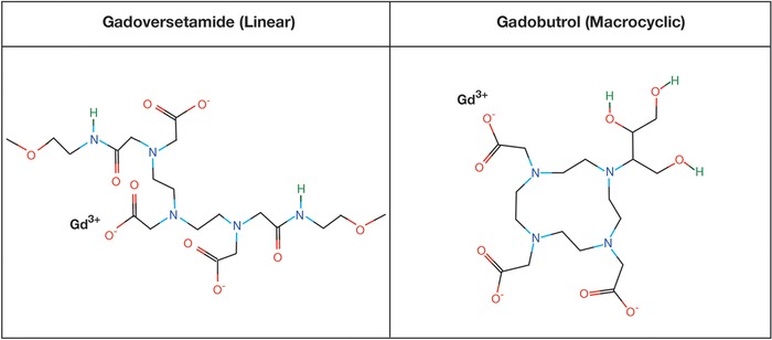
Background: Magnetic resonance imaging (MRI) enhanced with gadolinium-based contrast agents (GBCAs) is an essential tool in the diagnosis and management of many neurologic diseases, including multiple sclerosis, brain tumors, and infections. The clinical utility of GBCAs is evidenced by their widespread use. GBCAs are produced in macrocyclic and linear forms. Since 2014, evidence has suggested that repeated administration of GBCAs can lead to gadolinium deposition in the brain. Methods: We review the literature on gadolinium deposition, including both animal and human studies, as well as the literature on GBCA-associated health outcomes. Additionally, we summarize and discuss the updated medical society recommendations and perspectives on GBCA use in clinical practice. Results: The first publication reporting gadolinium deposition in the human brain was published in 2014. Since that seminal report, multiple studies have demonstrated that exposure to linear GBCAs is associated with gadolinium deposition in the dentate nucleus and globus pallidus as seen on brain MRI. Macrocyclic GBCA exposure has not convincingly been associated with gadolinium deposition evident on brain MRI. Conclusion: Clear evidence demonstrates that GBCAs lead to gadolinium deposition in the brain in a dose-dependent manner; however, only linear GBCAs have been associated with gadolinium deposition visualized on MRI. To date, no evidence links gadolinium deposition with any adverse health outcome. Updated medical society guidelines emphasize the importance of an individualized risk-benefit analysis with each administration of GBCAs.
Keywords: Contrast media; gadolinium; magnetic resonance imaging.
Figures
References
-
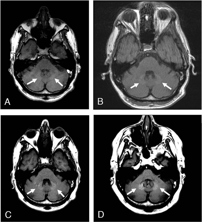
- Stojanov DA, Aracki-Trenkic A, Vojinovic S, Benedeto-Stojanov D, Ljubisavljevic S. Increasing signal intensity within the dentate nucleus and globus pallidus on unenhanced T1W magnetic resonance images in patients with relapsing-remitting multiple sclerosis: correlation with cumulative dose of a macrocyclic gadolinium-based contrast agent, gadobutrol. Eur Radiol. 2016. March;26(3):807-815. doi: 10.1007/s00330-015-3879-9. - DOI - PubMed
-
- Tweedle MF, Wedeking P, Kumar K. Biodistribution of radiolabeled, formulated gadopentetate, gadoteridol, gadoterate, and gadodiamide in mice and rats. Invest Radiol. 1995;30(6):372-380. - PubMed
-
- Kanda T, Ishii K, Kawaguchi H, Kitajima K, Takenaka D. High signal intensity in the dentate nucleus and globus pallidus on unenhanced T1-weighted MR images: relationship with increasing cumulative dose of a gadolinium-based contrast material. Radiology. 2014. March;270(3):834-841. doi: 10.1148/radiol.13131669. - DOI - PubMed
Publication types
LinkOut - more resources
Full Text Sources
Other Literature Sources