Review
. 2019 Mar 28:10:525.
doi: 10.3389/fmicb.2019.00525. eCollection 2019.

Pathways Exploited by Flaviviruses to Counteract the Blood-Brain Barrier and Invade the Central Nervous System

Affiliations
Review

Pathways Exploited by Flaviviruses to Counteract the Blood-Brain Barrier and Invade the Central Nervous System

Yasmin Mucunã Mustafá et al. Front Microbiol. .

Abstract

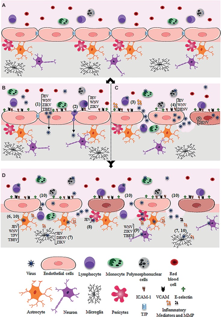
Human infection by different flaviviruses may cause severe neurologic syndromes, through pathogenic mechanisms that are still largely unknown. Japanese encephalitis virus (JEV), West Nile virus (WNV), Zika virus (ZIKV), yellow fever virus (YFV), dengue virus (DENV), and tick-borne encephalitis virus (TBEV) are believed to reach the central nervous system by a hematogenous route, upon crossing the blood-brain barrier. Although the disruption of BBB during flavivirus infection has been largely evidenced in experimental models, the relevance of BBB breakdown for virus entering the brain was not completely elucidated. In vitro models of BBB had demonstrated that these viruses replicated in brain microvascular endothelial cells (BMECs), which induced downregulation of tight junction proteins and increased the permeability of the barrier. Other reports demonstrated that infection of BMECs allowed the basolateral release of infectious particles, without a remarkable cytopathic effect, what might be sufficient for virus invasion. Virus replication and activation of other cells associated to the BBB, mostly astrocytes and microglia, were also reported to affect the endothelial barrier permeability. This event might occur simultaneously or after BMECs infection, being a secondary effect leading to BBB disruption. Importantly, activation of BMECs, astrocytes, and microglia by flaviviruses was associated to the expression and secretion of inflammatory mediators, which are believed to recruit leukocytes to the CNS. The leukocyte infiltrate could further mediate viral invasion through a Trojan horse mechanism and might contribute to BBB breakdown and to neurological alterations. This review discussed the previous studies regarding in vitro and in vivo models of JEV, WNV, ZIKV, YFV, DENV, and TBEV infection and addressed the pathways for BBB overcome and invasion of the CNS described for each virus infection, aiming to increment the knowledge and stimulate further discussion about the role of BBB in the neuropathogenesis of flavivirus infection.

Keywords: Japanese encephalitis virus; West Nile virus; Zika virus; blood-brain barrier; brain microvascular endothelial cells; dengue virus; flavivirus; yellow fever virus.


Figures

Figure 1
Figure 1
Schematic figure showing possible mechanism of virus entry into the central nervous system through the blood-brain barrier. (A) Intact blood-brain barrier is composed by endothelial cell strongly adhered through tight junction proteins (TJP), in association to pericytes, astrocytes, and microglia. The barrier controls the flux of solutes, blood cells (lymphocytes, monocytes, and polymorphonuclear cells), and pathogens from the blood to the central nervous system. (B) After systemic infection, some flavivirus reaches the BBB through hematogenous route and may cross the endothelial barrier without remarkable cytopathic effect. JEV, WNV, ZIKV, and TBEV may cross the endothelial barrier as cell-free virus (1). JEV, WNV, and ZIKV were also reported to traverse the endothelial barrier associated to infected leukocytes (2). (C) Systemic infection and inflammation and/or the direct infection of brain endothelial cells may induce BBB breakdown, allowing virus invasion of the CNS. Regarding this, systemic inflammation due to activation of immune cells upon infection was associated to the release of inflammatory mediators, which then affect the permeability of the endothelial barrier (3). Also, replication of JEV, WNV, and DENV in the brain endothelial cells may induce downregulation of TJP expression (4) and/or cell death (5), promoting the barrier disruption and virus entry. (D) After entry into the brain by any of the described pathways (B and C), flaviviruses may infect the astrocytes (as described for JEV, WNV, YFV, TBEV) (6), microglia (as described for JEV, DENV, ZIKV) (7), pericytes (as described for JEV) (8), and neurons (as described for WNV, ZIKV, TBEV) (9). The infection of these cells, especially astrocytes and microglia, induces the release of inflammatory mediators (IL-6, VEGF, TNF-α, IFN-γ, IL-1β and IL-10, MCP-1) and metalloproteinases (MMP2, MMP3, MM9) (10), which mediate the downregulation of adherents and tight junction proteins (TJP), resulting in increased permeability.

References

    1. Agrawal T., Sharvani V., Nair D., Medigeshi G. R. (2013). Japanese encephalitis virus disrupts cell-cell junctions and affects the epithelial permeability barrier functions. PLoS One 8, 1–16. 10.1371/journal.pone.0069465 - DOI - PMC - PubMed
    1. Alimonti J. B., Ribecco-Lutkiewicz M., Sodja C., Jezierski A., Stanimirovic D. B., Liu Q., et al. (2018). Zika virus crosses an in vitro human blood brain barrier model. Fluids Barriers CNS 15, 1–9. 10.1186/s12987-018-0100-y - DOI - PMC - PubMed
    1. Al-Obaidi M. M. J., Bahadoran A., Har L. S., Mui W. S., Rajarajeswaran J., Zandi K., et al. . (2017). Japanese encephalitis virus disrupts blood-brain barrier and modulates apoptosis proteins in THBMEC cells. Virus Res. 233, 17–28. 10.1016/j.virusres.2017.02.012, PMID: - DOI - PubMed
    1. Alvarez J. I., Katayama T., Prat A. (2013). Glial influence on the blood brain barrier. Glia 61, 1939–1958. 10.1002/glia.22575, PMID: - DOI - PMC - PubMed
    1. Araújo F. M., Brilhante R. S., Cavalcanti L. P., Rocha M. F., Cordeiro R. A., Perdigão A. C., et al. . (2011). Detection of the dengue non-structural 1 antigen in cerebral spinal fluid samples using a commercial ayavailable enzyme-linked immunosorbent assay. J. Virol. Methods 177, 128–131. 10.1016/j.jviromet.2011.07.003, PMID: - DOI - PubMed

LinkOut - more resources