. 2019 Mar 20:10:536.
doi: 10.3389/fimmu.2019.00536. eCollection 2019.

Defective Induction of COX-2 Expression by Psoriatic Fibroblasts Promotes Pro-inflammatory Activation of Macrophages

Affiliations

Defective Induction of COX-2 Expression by Psoriatic Fibroblasts Promotes Pro-inflammatory Activation of Macrophages

Jorge Arasa et al. Front Immunol. .

Abstract

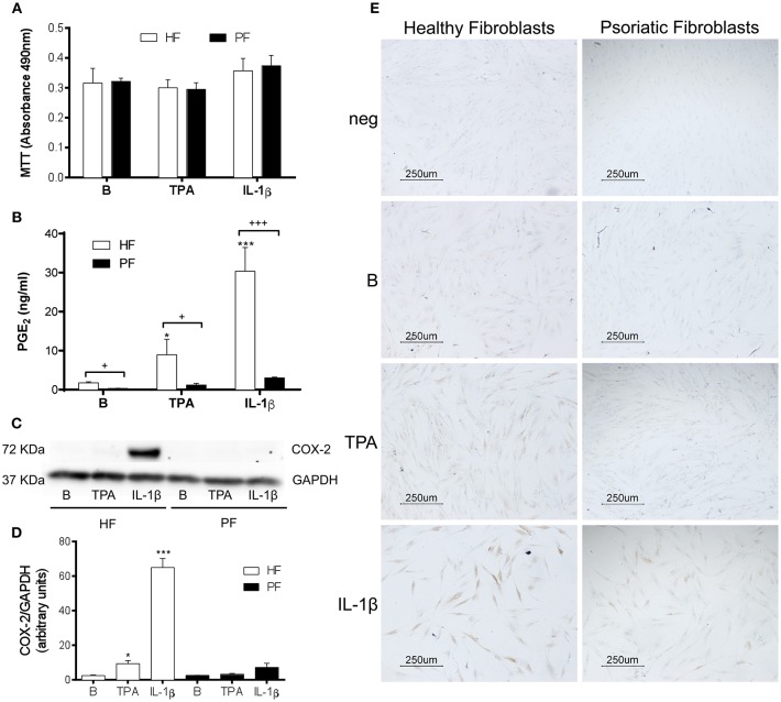
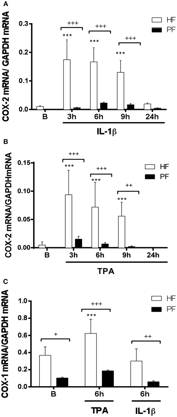
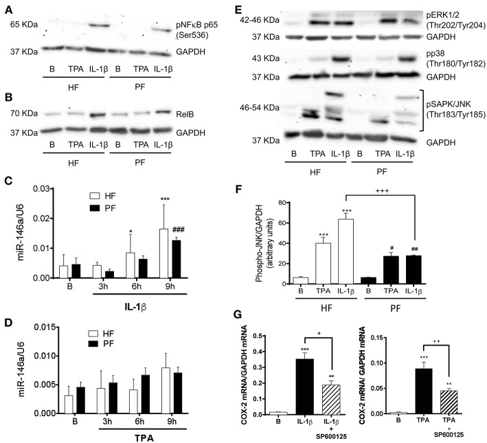
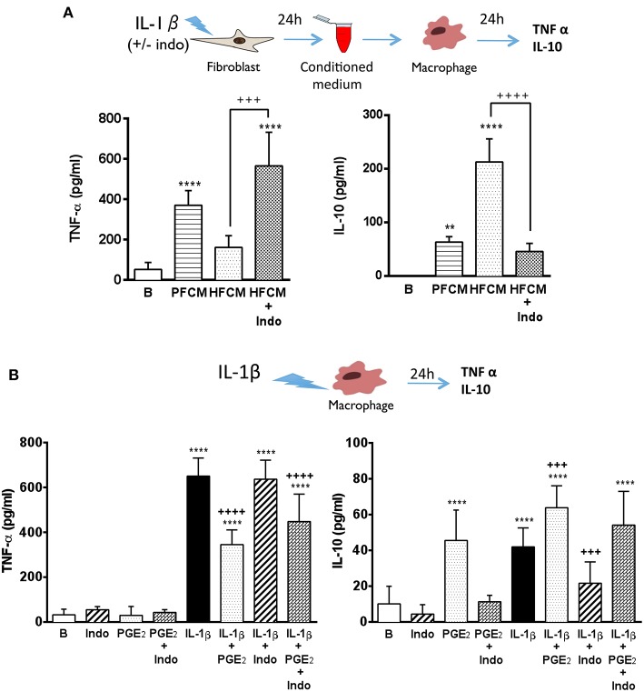
Fibroblasts play an important role as members of the innate immune system through the secretion of COX-2-derived inflammatory mediators such as prostaglandin E2 (PGE2). However, it has been described that dermal fibroblasts behave like mesenchymal stem cells reducing lymphocyte recruitment and dendritic cell activation through PGE2 release. As the role of fibroblasts in psoriasis remains poorly characterized, in the present study we have evaluated the possible influence of PGE2 derived from dermal fibroblasts as modulator of the immune response in psoriatic skin. Our results indicate that under inflammatory conditions, psoriatic fibroblasts showed defective induction of COX-2, which resulted in diminished production of PGE2, in contrast to healthy fibroblasts. This phenotype correlated with deficient c-Jun N-terminal kinase (JNK) activation, in accordance with the hypothesis that alterations in members of the JNK pathway are associated with psoriasis. Furthermore, conditioned medium from psoriatic fibroblasts promoted the polarization of monocytic cells toward a pro-inflammatory profile, effect that was mimicked in healthy fibroblasts after pre-incubation with indomethacin. These results are consistent with a prominent role of dermal fibroblasts in the regulation of inflammatory response through the participation of COX-derived metabolites. This resolutive behavior seems to be defective in psoriatic fibroblasts, offering a possible explanation for the chronification of the disease and for the exacerbation triggered by nonsteroidal anti-inflammatory drugs (NSAIDS) such as indomethacin.

Keywords: cyclooxygenase; fibroblasts; inflammation; macrophages; psoriasis.


Figures

Figure 1
Figure 1
PGE2 production and COX-2 expression are decreased in stimulated psoriatic fibroblasts. Cells were treated with 2.5 ng/ml IL-1β or 1 μg/ml TPA for 24 h. (A) 3-(4,5-dimethylthiazol-2-yl)-2,5-diphenyltetrazolium bromide (MTT) assay for cell viability (n = 4 biopsies) and (B) Prostaglandin E2 (PGE2) determined by radioimmunoassay (n = 6 biopsies) were performed in duplicate. (C,D) COX-2 protein expression assessed by Western blotting (n = 3 biopsies). Data represent mean ± SD. *p < 0.05, ***p < 0.001 vs. unstimulated fibroblasts (B) and +p < 0.05, +++p < 0.001 vs. healthy fibroblasts (HF) using Sidak's multiple comparison test. PF, psoriatic fibroblasts. (E) COX-2 protein expression determined by immunocytochemistry (representative photomicrographs of three independent experiments).
Figure 2
Figure 2
COX-1 and COX-2 mRNA expression is decreased in psoriatic fibroblasts. Cells were treated with 2.5 ng/ml IL-1β (A) or 1 μg/ml TPA (B) for 3, 6, 9, or 24 h and COX-2 mRNA levels were evaluated by quantitative real-time PCR. (C) COX-1 mRNA levels were evaluated by quantitative PCR after IL-1β or TPA stimulation during 6 h. Data represent mean ± SD (n = 6 biopsies) of mRNA expression normalized to the housekeeping gene GAPDH and expressed as 2−ΔΔCT values. ***p < 0.001 vs. unstimulated healthy fibroblasts (B) and +p < 0.05, ++p < 0.01, +++p < 0.001 vs. healthy fibroblasts (HF) using Sidak's multiple comparison test. PF, psoriatic fibroblasts.
Figure 3
Figure 3
Psoriatic fibroblasts show an altered activation of the SAPK/JNK pathway involved in COX-2 induction. (A) Representative immunoblotting image of phosphorylated NF-κB p65 subunit after 15 min treatment with 2.5 ng/ml IL-1β or 1 μg/ml TPA. (B) Representative immunoblotting image of phosphorylated NF-κB RelB subunit after 6 h treatment with IL-1β or TPA. (C,D) miR146a expression determined by RT-qPCR at 0, 3, 6, or 9 h post stimulation with IL-1β or TPA. Data represent mean ± SD (n = 4 biopsies) of 2−ΔΔCT values normalized to the small nucleolar RNA U6. *p < 0.05, ***p < 0.001 vs. non stimulated (B) healthy fibroblasts (HF) and ###p < 0.001 vs. non stimulated (B) psoriatic fibroblasts (PF) using Sidak's multiple comparison test. (E) Representative immunoblotting image of phosphorylated p38, ERK1/2, and SAPK/JNK after 15 min stimulation with IL-1β or TPA. (F) Densitometry analysis of phospho SAPK/JNK immunoblots. Data represent mean ± SD (n = 4). ***p < 0.001 vs. non stimulated (B) healthy fibroblasts (HF); #p < 0.05, ##p < 0.01 vs. non stimulated (B) psoriatic fibroblasts (PF) and +++ p < 0.001 vs. healthy fibroblasts (HF) using Sidak's multiple comparison test. (G) RT-qPCR for COX-2 mRNA in HF after preincubation with the JNK inhibitor SP600125 (50 μM) during 45 min and the subsequently stimulation with IL-1β (n = 6 biopsies) or TPA (n = 8 biopsies) during 2 h. Data represent mean ± SD. **p < 0.01, ***p < 0.001 vs. unstimulated cells (B); +p < 0.05, ++p < 0.01 vs. IL-1β/TPA stimulated fibroblasts using Tukey's multiple comparison test.
Figure 4
Figure 4
Psoriatic fibroblasts support pro-inflammatory activation of macrophages. (A) TNF-α and IL-10 levels in THP-1 derived macrophages incubated during 24 h with conditioned media from IL-1β-stimulated psoriatic fibroblast (PFCM) (n = 4 biopsies) or IL-1β-stimulated healthy fibroblasts (HFCM) (n = 4 biopsies). When indicated, healthy fibroblasts where pretreated with 10 μM indomethacin (Indo) before stimulation. Data represent mean ± SD. **p < 0.01, ****p < 0.0001 respect untreated macrophages (B); +++p < 0.001 ++++p < 0.0001 vs. macrophages treated with HFCM using Tukey's multiple comparison test. (B) TNF-α and IL-10 levels in THP-1 derived macrophages treated during 24 h with PGE2 (30 ng/ml), indomethacin (10 μM) or their combination in the presence or the absence of IL-1β (2.5 ng/ml). Data represent mean ± SD (n = 6). ***p < 0.001, ****p < 0.0001 vs. untreated cells (B). +++p < 0.001 respect IL-1β-treated cells using Tukey's multiple comparison test. All conditions were assayed in duplicate.

References

    1. Girolomoni G, Strohal R, Puig L, Bachelez H, Barker J, Boehncke WH, et al. The role of IL-23 and the IL-23/TH17 immune axis in the pathogenesis and treatment of psoriasis. J Eur Acad Dermatol Venereol. (2017) 31:1616–26. 10.1111/jdv.14433 - DOI - PMC - PubMed
    1. Di Meglio P, Villanova F, Nestle FO. Psoriasis. Cold Spring Harb Perspect Med. (2014) 4:a015354. 10.1101/cshperspect.a015354 - DOI - PMC - PubMed
    1. Lowes MA, Suarez-Farinas M, Krueger JG. Immunology of psoriasis. Annu Rev Immunol. (2014) 32:227–55. 10.1146/annurev-immunol-032713-120225 - DOI - PMC - PubMed
    1. Perera GK, Di Meglio P, Nestle FO. Psoriasis. Annu Rev Pathol-Mech. (2012) 7:385–422. 10.1146/annurev-pathol-011811-132448 - DOI - PubMed
    1. Buckley CD. Why should rheumatologists care about fibroblasts?: Answering questions about tissue tropism and disease persistence. Rheumatology. (2017) 56:863–4. 10.1093/rheumatology/kew289 - DOI - PubMed

Publication types