Review
. 2019 Mar 22:10:553.
doi: 10.3389/fimmu.2019.00553. eCollection 2019.

IgA and FcαRI: Pathological Roles and Therapeutic Opportunities

Affiliations
Review

IgA and FcαRI: Pathological Roles and Therapeutic Opportunities

Annelot Breedveld et al. Front Immunol. .

Abstract

Immunoglobulin A (IgA) is the most abundant antibody class present at mucosal surfaces. The production of IgA exceeds the production of all other antibodies combined, supporting its prominent role in host-pathogen defense. IgA closely interacts with the intestinal microbiota to enhance its diversity, and IgA has a passive protective role via immune exclusion. Additionally, inhibitory ITAMi signaling via the IgA Fc receptor (FcαRI; CD89) by monomeric IgA may play a role in maintaining homeostatic conditions. By contrast, IgA immune complexes (e.g., opsonized pathogens) potently activate immune cells via cross-linking FcαRI, thereby inducing pro-inflammatory responses resulting in elimination of pathogens. The importance of IgA in removal of pathogens is emphasized by the fact that several pathogens developed mechanisms to break down IgA or evade FcαRI-mediated activation of immune cells. Augmented or aberrant presence of IgA immune complexes can result in excessive neutrophil activation, potentially leading to severe tissue damage in multiple inflammatory, or autoimmune diseases. Influencing IgA or FcαRI-mediated functions therefore provides several therapeutic possibilities. On the one hand (passive) IgA vaccination strategies can be developed for protection against infections. Furthermore, IgA monoclonal antibodies that are directed against tumor antigens may be effective as cancer treatment. On the other hand, induction of ITAMi signaling via FcαRI may reduce allergy or inflammation, whereas blocking FcαRI with monoclonal antibodies, or peptides may resolve IgA-induced tissue damage. In this review both (patho)physiological roles as well as therapeutic possibilities of the IgA-FcαRI axis are addressed.

Keywords: CD89; IgA; IgA deficiency; autoimmunity; microbiome; mucosa; therapy; vaccination.


Figures
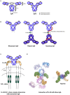
Figure 1
Figure 1
Structure of IgA isotypes and the IgA Fc receptor (FcαRI). (A) IgA1 vs. IgA2. IgA consists of two heavy chains (blue), each composed of three constant regions and one variable region, and two light chains (brown) that consist of one constant and one variable region. IgA1 contains a hinge region with O-linked glycosylation. (B) Monomeric, dimeric IgA, and secretory IgA. Dimeric IgA consist of two IgA molecules that are linked with a J-chain (green). Secretory IgA contains an additional molecule, the secretory component (SC; red). (C) Structure of FcαRI. FcαRI consist of a transmembrane domain, a short cytoplasmic tail and two extracellular domains (EC1 & EC2). It is associated with the signaling FcR γ chain via an electrostatic interaction. The IgA heavy chain junction of Cα2 and Cα3 binds to the EC1 domain of FcαRI in a 2:1 stoichiometry. (D) A model of dIgA1 bound to four FcαRI molecules. The FcαRI:IgA1-Fc complex (PDB 1OW0) was superimposed onto the solution structure of dIgA1 published by Bonner et al. (24) (PDB 2QTJ). FcαRI molecules bound to the top or bottom IgA1 antibodies are colored green or yellow, respectively. The C-terminal residue of each receptor is shown in orange or red to illustrate the membrane-proximal region. Kindly provided by Andrew B. Herr, PhD (Cincinnati Children's Hospital).
Figure 2
Figure 2
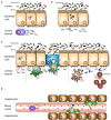
Roles of mucosal IgA in homeostasis. (A) Dimeric IgA (dIgA) is produced by local plasma cells (PC) in the lamina propria. Dimeric IgA is transported to the intestinal lumen by binding to the polymeric immunoglobulin receptor (pIgR) present on epithelial cells. In the lumen it is released as secretory IgA (SIgA) where it can coat (commensal) bacteria. (B) On route dIgA can bind, neutralize and eliminate viruses. (C) (1) Infiltrated antigens and pathogens are opsonized by dIgA and transported back into the lumen. Sub epithelial dendritic cells (DCs) can (2) sample antigens or (3) take up SIgA-coated pathogens that enter via microfold cells (M cells). Pathogens in the lamina propria are coated with dIgA after which this immune complex is taken up by FcαRI- expressing (4) DCs, and (5) neutrophils. In response neutrophils secrete leukotriene B4 (LTB4), hereby attracting more neutrophils, which will clear the infection. (D) Serum IgA is (1) capable of inhibiting (unwanted) pro inflammatory responses in the circulation via ITAMi signaling in monocytes. (2) IgA-opsonized bacteria which have leaked into the circulation are taken up by FcαRI-expressing Kupffer cells (KC) in the liver.
Figure 3
Figure 3
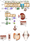
Mucosal IgA in pathogenesis. (A) IgA deficiency. In absence of IgA the intestinal microbiota is (1) less diverse and unbalanced. (2) IgM, and (3) IgG can be transported across the epithelium by associating with the pIgR or the neonatal Fc receptor (FcRn), respectively, to compensate IgA loss. Theoretically, IgM may be able to (4) neutralize viruses and (5) exclude infiltrated antigens. Sub epithelial DCs are not described to (6) sample IgM coated bacteria. (7) IgM-coated bacteria are not described to enter M cells. (8) Invaded IgM-coated pathogens are not recognized by FcαRI-expressing immune cells, resulting in less efficient pathogen elimination. (B) Celiac disease and gluten-associated diseases. (1) IgA-gliadin complexes bind to the transferrin receptor (TfR) and are retrotransported across the epithelium. (2) In the lamina propria deamidated gliadin is taken up by DCs and after processing (3) presented to T helper (Th) cells. (4) Th1 cell activation leads to the release of pro-inflammatory mediators (5) causing tissue damage. (6) Activated Th2 cells stimulate autoantibody production directed against gliadin and tissue transglutaminase by B cells. (7) Plasma cells produce autoantibodies which (8) can be detected in the circulation and form deposits with soluble FcαRI in the kidney resulting in damage (IgA nephropathy). (9) IgA anti-tissue transglutaminase forms complexes in the dermis, resulting in neutrophil activation and concomitant tissue damage (dermatitis herpetiformis). (C) IgA-FcαRI induced pathology. IgA immune complexes activate neutrophils by cross-linking FcαRI, resulting in release of LTB4 and enhanced neutrophil influx, which induces tissue damage as seen in the skin (LABD), vessels (IgA vasculitis), joints (RA), and potentially in colon (IBD).
Figure 4
Figure 4
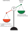
IgA and FcαRI as therapeutic targets. (1) Enhanced IgA-FcαRI activation in IgA-associated autoimmune diseases is unwanted. Blocking IgA-FcαRI interactions by monoclonal antibodies or peptides may reduce tissue damage in these diseases. (2) Treatment with monomeric IgA or anti-FcαRI Fabs may induce ITAMi signaling thereby inhibiting IgG-induced phagocytosis and IgE-mediated allergic diseases. (3) To combat infections, administration of IgA (passive vaccination) or induction of IgA levels via active vaccination may result in enhanced protective immunity. (4) IgA monoclonal antibody therapy may result in efficient killing of tumor cells by activating FcαRI-expressing immune cells.

References

    1. Woof JM, Kerr MA. The function of immunoglobulin A in immunity. J Pathol. (2006) 208:270–82. 10.1002/path.1877 - DOI - PubMed
    1. Vidarsson G, Dekkers G, Rispens T. IgG subclasses and allotypes: from structure to effector functions. Front Immunol. (2014) 5:520. 10.3389/fimmu.2014.00520 - DOI - PMC - PubMed
    1. Wines BD, Hogarth PM. IgA receptors in health and disease. Tissue Antigens. (2006) 68:103–14. 10.1111/j.1399-0039.2006.00613.x - DOI - PubMed
    1. Macpherson AJ, Yilmaz B, Limenitakis JP, Ganal-Vonarburg SC. IgA function in relation to the intestinal microbiota. Annu Rev Immunol. (2018) 36:359–81. 10.1146/annurev-immunol-042617-053238 - DOI - PubMed
    1. Leong KW, Ding JL. The unexplored roles of human serum IgA. DNA Cell Biol. (2014) 33:823–9. 10.1089/dna.2014.2639 - DOI - PMC - PubMed

Publication types