. 2019 Mar 5;9(4):743-754.
doi: 10.1002/2211-5463.12613. eCollection 2019 Apr.

Tocopherol biosynthesis in Leishmania (L.) amazonensis promastigotes

Affiliations

Tocopherol biosynthesis in Leishmania (L.) amazonensis promastigotes

José Mário F Balanco et al. FEBS Open Bio. .

Abstract

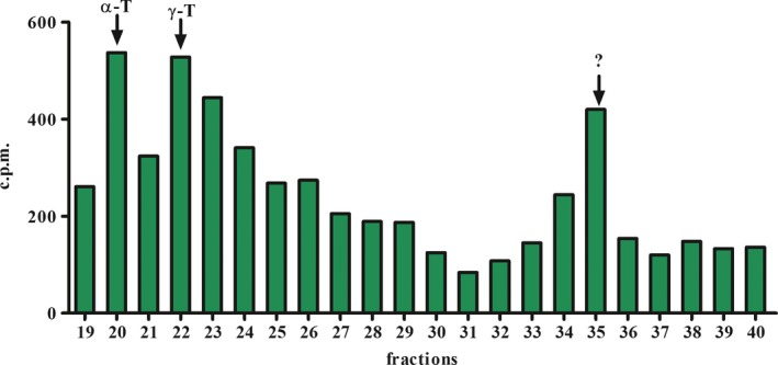
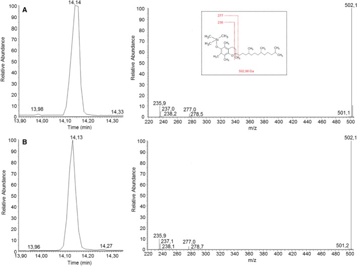
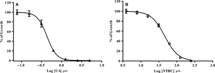
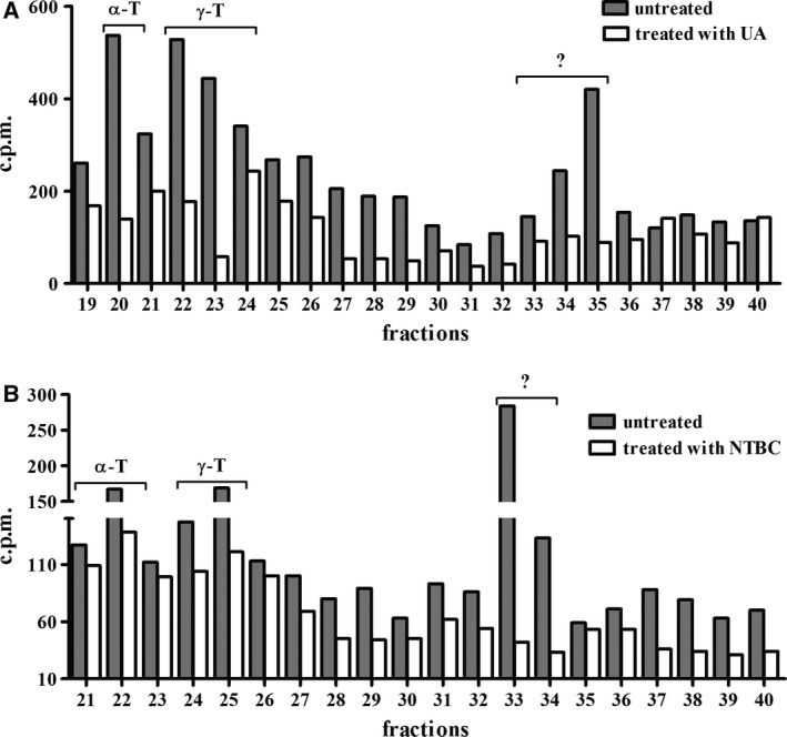
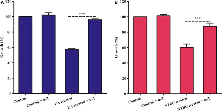
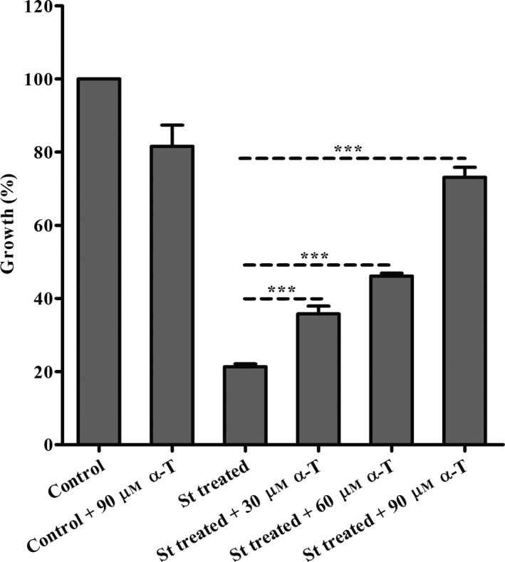
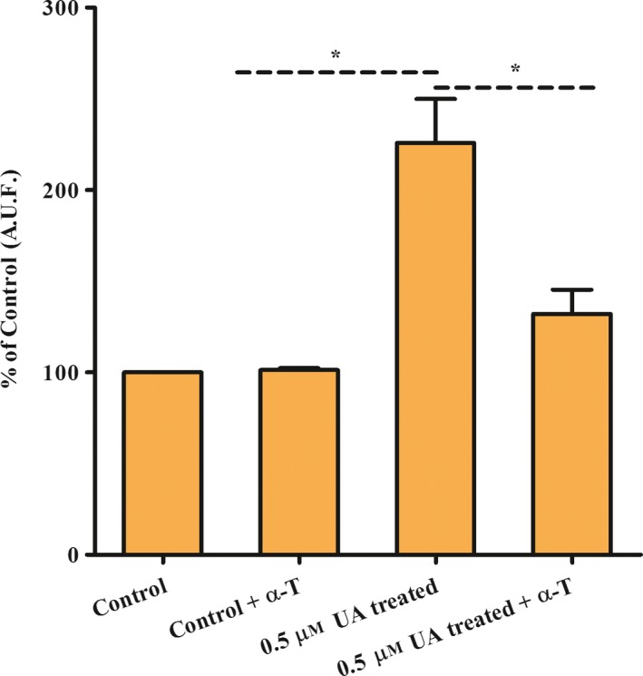
Leishmaniasis is a neglected disease caused by a trypanosomatid protozoan of the genus Leishmania. Most drugs used to treat leishmaniasis are highly toxic, and the emergence of drug-resistant strains has been observed. Therefore, new therapeutic targets against leishmaniasis are required. Several isoprenoid compounds, including dolichols or ubiquinones, have been shown to be important for cell viability and proliferation in various trypanosomatid species. Here, we detected the biosynthesis of tocopherol in Leishmania (L.) amazonensis promastigotes in vitro through metabolic labelling with [1-(n)-3H]-phytol. Subsequently, we confirmed the presence of vitamin E in the parasite by gas chromatography-mass spectrometry. Treatment with usnic acid or nitisinone, inhibitors of precursors of vitamin E synthesis, inhibited growth of the parasite in a concentration-dependent manner. This study provides the first evidence of tocopherol biosynthesis in a trypanosomatid and suggests that inhibitors of the enzyme 4-hydroxyphenylpyruvate dioxygenase may be suitable for use as antileishmanial compounds.

Database: The amino acid sequence of a conserved hypothetical protein [Leishmania mexicana MHOM/GT/2001/U1103] has been deposited in GenBank (CBZ28005.1).

Keywords: Leishmania (L.) amazonensis; isoprenoid; nitisinone, tocopherol; trypanosomatid; usnic acid.


Conflict of interest statement

The authors declare no conflict of interest.

Figures

Figure 1
Figure 1
Radioactive elution profile employing radiolabelled precursor and/or RPHPLC system. Profile of the promastigotes of Leishmania (L.) amazonensis, metabolically labelled with [1‐(n)‐3H]‐phytol. Extracts from promastigotes stages were purified by RPHPLC. Fractions were collected at intervals of 1 mL·min−1. The retention time of compounds was identified by co‐injection of commercial standards in both systems. α‐T: α‐tocopherol, γ‐T: γ‐tocopherol. ?: unknown. Three independent experiments. c.p.m., count per minute.
Figure 2
Figure 2
Molecular identification of α‐tocopherol biosynthesis in promastigotes of Leishmania (L.) amazonensis by GCMS/MS. Chromatograms and spectra from (A) α‐tocopherol standard; (B) HPLC peaks from 1.25 × 1010 parasites. All samples were previously purified by RPHPLC. The molecular structure was confirmed by comparing the retention time of GC and the MS2 spectrum of the parental ions at m/z 502 for α‐tocopherol.
Figure 3
Figure 3
Dose–response curves of parasite viability in the presence of usnic acid (UA) or nitisinone (NTBC) at 72 h of treatment by MTT methodology. (A) Usnic acid inhibited parasite growth in a dose‐dependent manner (IC 50: 0.433 ± 0.071 μm), and (B) NTBC inhibited parasite growth in a dose‐dependent manner too (IC 50: 47.87 ± 10 μm). Data are representative of three independent experiments; each experiment done contained three technical triplicates. These data are expressed as means ± SD.
Figure 4
Figure 4
Inhibition of vitamin E biosynthesis in parasites treated with UA or NTBC for in vitro cultures of the Leishmania (L.) amazonensis promastigotes. (A) Radioactive elution profile of promastigotes treated (white bars) or untreated (grey bars) for 72 h with UA and labelled with [1‐(n)‐3H]‐phytol in the last 24 h. α‐T: α‐tocopherol, γ‐T: γ‐tocopherol, ?: unknown. (B) Radioactive elution profile of promastigotes treated (white bars) or untreated (grey bars) for 48 h with NTBC and labelled with [1‐(n)‐3H]‐phytol in the last 24 h. α‐T: α‐tocopherol, γ‐T: γ‐tocopherol, ?: unknown. Data are representative of two independent experiments. c.p.m.: count per minute.
Figure 5
Figure 5
Effect of α‐tocopherol exogenous in recover of growth of promastigotes treated with UA or NTBC. (A) Recovery of parasites treated for 72 h at 25 °C with UA by exogenously added α‐tocopherol. (Control) parasites control; (control + α‐T) parasites control plus α‐tocopherol; (UA treated) parasites treated with 0.5 μm of UA; (UA treated plus α‐T) parasites treated with 0.5 μm of UA plus 25 μm of α‐tocopherol. (B) Recovery of parasites treated for 48 h at 25 °C with NTBC by α‐tocopherol exogenous. (Control) parasites control; (control + α‐T) parasites control plus α‐tocopherol; (NTBC treated) parasites treated with 31.25 μm of NTBC; (NTBC treated + α‐T) parasites treated with 31.25 μm of NTBC plus 200 μm α‐tocopherol. O.D., optical density. MTT for 1 h at 25 °C. (***) P < 0.0001 ANOVA. Data are representative of three independent experiments; each experiment done contained three technical triplicates. These data are expressed as means ± SD.
Figure 6
Figure 6
Recovery test of Leishmania (L.) amazonensis promastigotes treated with staurosporine (St) and α‐tocopherol (α‐T) at 25 °C for 72 h. (Control) parasites control; (control + 90 μm α‐T) parasites control plus 90 μm of α‐tocopherol; (St treated) parasites treated with 75 nm staurosporine; (St treated + 30 μm α‐T) parasites treated with 75 nm staurosporine plus 30 μm α‐tocopherol; (St treated + 60 μm α‐T) 75 nm staurosporine plus 60 μm α‐tocopherol; (St treated + 90 μm α‐T) parasites treated with 75 nm staurosporine plus 90 μm α‐tocopherol. Viability by MTT assay for 1 h at 25 °C. OD, optical density. (***) P < 0.0001 ANOVA. Data are representative of three independent experiments; each experiment done contained three technical triplicates. These data are expressed as means ± SD.
Figure 7
Figure 7
Reactive oxygen species (ROS) measurement in Leishmania (L.) amazonensis promastigotes UA treated for 96 h. CellROX assay I. (Control) parasites control; (control + α‐T) parasites plus α‐tocopherol; (0.5 μm UA treated) parasites treated with 0.5 μm of usnic acid and (0.5 μm UA treated + α‐T) parasites treated with 0.5 μm of usnic acid plus 25 μm of α‐tocopherol. ROS was estimated by the AUF (arbitrary unit of fluorescence). (*) P < 0.005 ANOVA. Data are representative of three independent experiments; each experiment done contained two technical triplicates. These data are expressed as means ± SD.

References

    1. Torres‐Guerrero E, Quintanilla‐Cedillo MR, Ruiz‐Esmenjaud J and Arenas R (2017) Leishmaniasis: a review. F1000Res 6, 750. - PMC - PubMed
    1. Trinconi CT, Reimao JQ, Coelho AC and Uliana SR (2016) Efficacy of tamoxifen and miltefosine combined therapy for cutaneous leishmaniasis in the murine model of infection with Leishmania amazonensis . J Antimicrob Chemother 71, 1314–1322. - PubMed
    1. Wang KC and Ohnuma S (2000) Isoprenyl diphosphate synthases. Biochim Biophys Acta 1529, 33–48. - PubMed
    1. Urbina JA (1997) Lipid biosynthesis pathways as chemotherapeutic targets in kinetoplastid parasites. Parasitology 114 (Suppl), S91–S99. - PubMed
    1. Pena‐Diaz J, Montalvetti A, Flores CL, Constan A, Hurtado‐Guerrero R, De Souza W, Gancedo C, Ruiz‐Perez LM and Gonzalez‐Pacanowska D (2004) Mitochondrial localization of the mevalonate pathway enzyme 3‐Hydroxy‐3‐methyl‐glutaryl‐CoA reductase in the Trypanosomatidae. Mol Biol Cell 15, 1356–1363. - PMC - PubMed

Publication types

MeSH terms