

Sclerosing pneumocytoma: Case report of a rare endobronchial presentation
- PMID: 30985653
- PMCID: PMC6485897
- DOI: 10.1097/MD.0000000000015038
Sclerosing pneumocytoma: Case report of a rare endobronchial presentation
Abstract
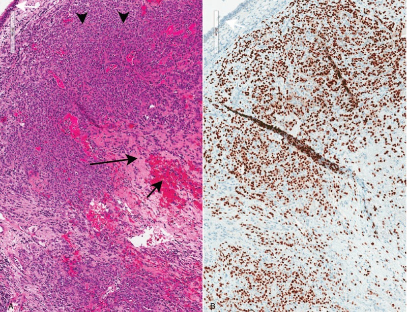
Rationale: Sclerosing pneumocytoma is a rare benign lung neoplasm seen in middle aged adults with a female predominance. Originally thought to be vascular in origin, this rare entity is now understood to be epithelial in nature. On imaging, sclerosing pneumocytoma manifests as a well circumscribed nodule or mass, often juxtapleural in location. On histopathology, sclerosing pneumocytoma is composed of cuboidal "surface cells" and round "stromal cells," both of which show nuclear staining for thyroid transcription factor-1 (TTF-1). Here we review the existing literature on sclerosing pneumocytoma and present a case of sclerosing pneumocytoma in a highly unusual endobronchial location.
Patient concerns: This case is a 43 year old woman who presented with chronic cough.
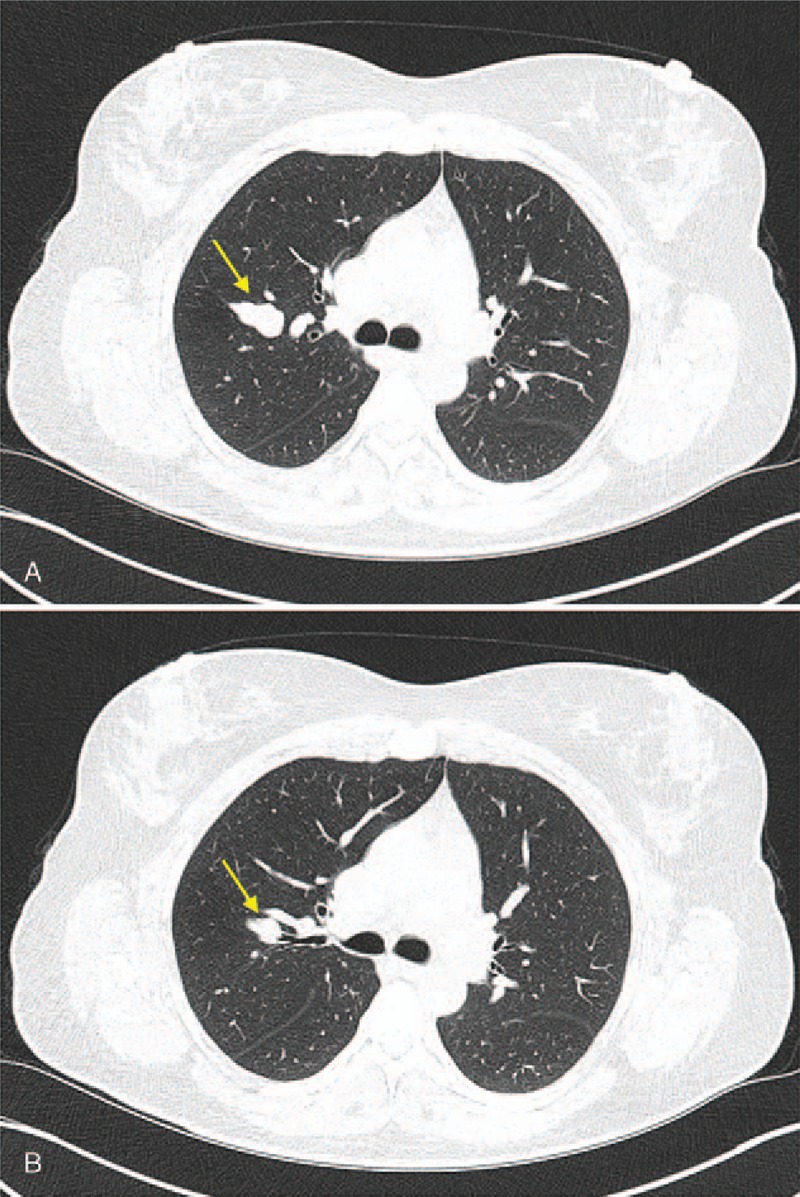
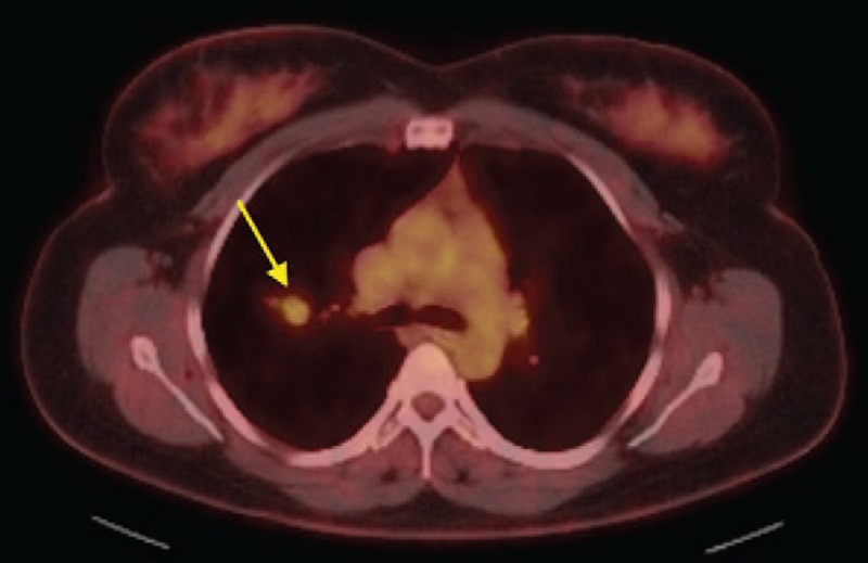
Diagnosis: Imaging revealed a right upper lobe nodule with an endobronchial component.
Interventions and outcomes: Endoscopic biopsy was performed, and pathologic diagnosis was confirmed.
Lessons: Although extremely rare, endobronchial presentation of sclerosing pneumocytoma is possible, and should remain on the differential for patients with endobronchial pulmonary lesions. Pathologic tissue analysis is necessary to confirm this uncommon diagnosis.
Conflict of interest statement
The authors have no conflicts of interest to disclose.
Figures
References
-
- Liebow AA, Hubbell DS. Sclerosing hemangioma (histiocytoma, xanthoma) of the lung. Cancer 1956;9:53–75. http://www.ncbi.nlm.nih.gov/pubmed/13284701http://www.ncbi.nlm.nih.gov/pubmed/13284701. Accessed March 24, 2018. - PubMed
-
- Devouassoux-Shisheboran M, Hayashi T, Linnoila RI, et al. A clinicopathologic study of 100 cases of pulmonary sclerosing hemangioma with immunohistochemical studies: TTF-1 is expressed in both round and surface cells, suggesting an origin from primitive respiratory epithelium. Am J Surg Pathol 2000;24:906–16. http://www.ncbi.nlm.nih.gov/pubmed/10895813http://www.ncbi.nlm.nih.gov/pubmed/10895813. Accessed March 14, 2018. - PubMed
-
- Travis WD, Brambilla E, Nicholson AG, et al. The 2015 World Health Organization classification of lung tumors: impact of genetic, clinical and radiologic advances since the 2004 classification. J Thorac Oncol 2015;10:1243–60. doi:10.1097/JTO.0000000000000630. - PubMed
-
- Im JG, Kim WH, Han MC, et al. Sclerosing hemangiomas of the lung and interlobar fissures: CT findings. J Comput Assist Tomogr 1994;181:34–8. http://www.ncbi.nlm.nih.gov/pubmed/8282879http://www.ncbi.nlm.nih.gov/pubmed/8282879. Accessed March 23, 2018. - PubMed
Publication types
MeSH terms
LinkOut - more resources
Full Text Sources
