Multicenter Study
. 2019 Aug 1;28(15):2531-2548.
doi: 10.1093/hmg/ddz075.

The protective variant rs7173049 at LOXL1 locus impacts on retinoic acid signaling pathway in pseudoexfoliation syndrome

Affiliations
Multicenter Study

The protective variant rs7173049 at LOXL1 locus impacts on retinoic acid signaling pathway in pseudoexfoliation syndrome

Daniel Berner et al. Hum Mol Genet. .

Abstract

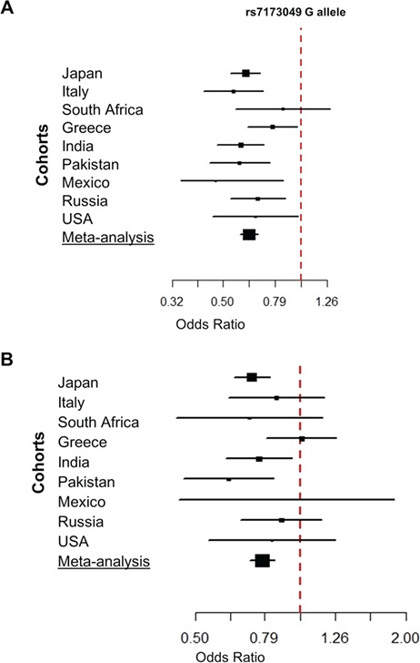
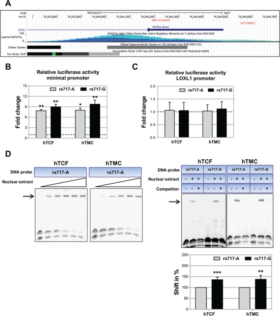
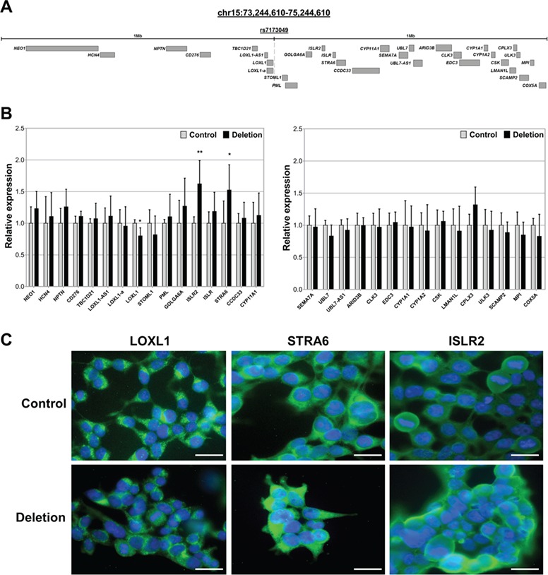
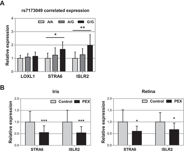
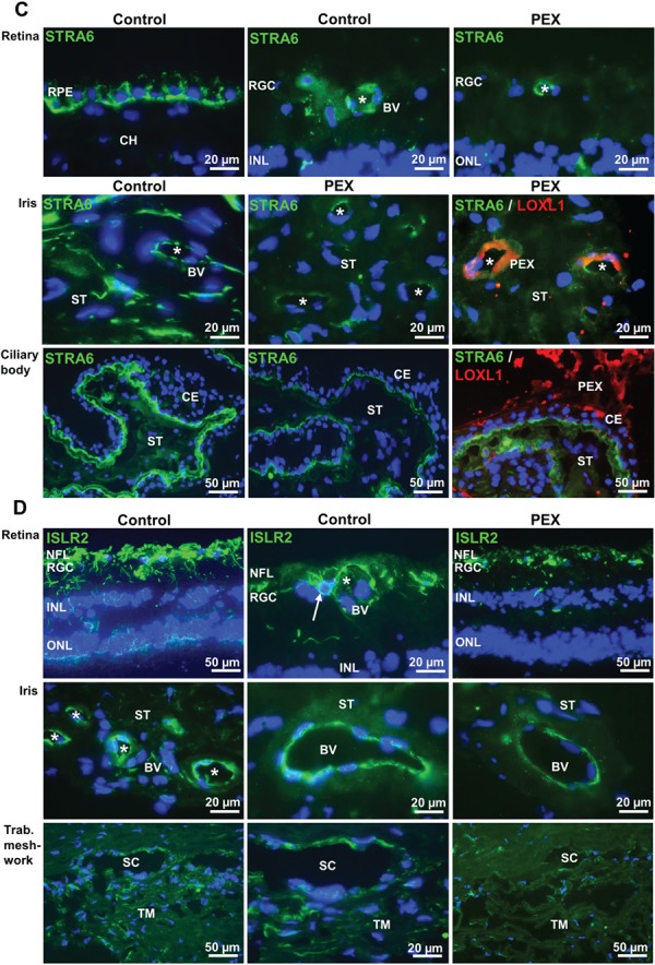
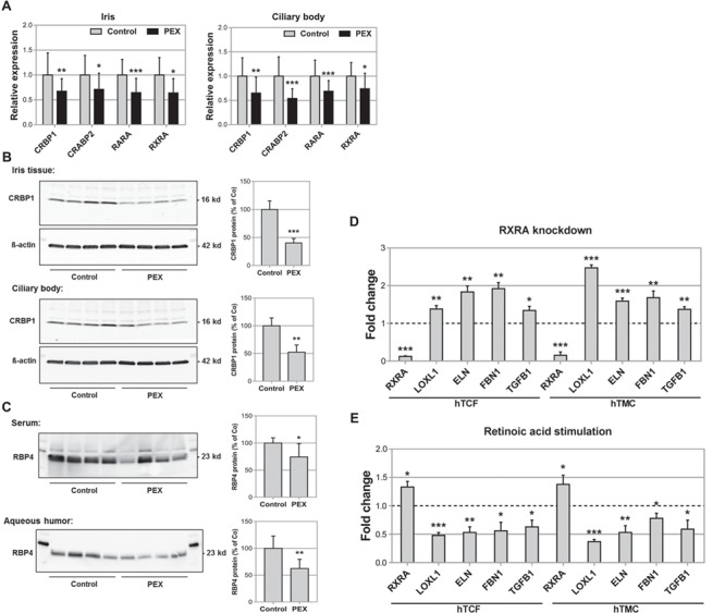
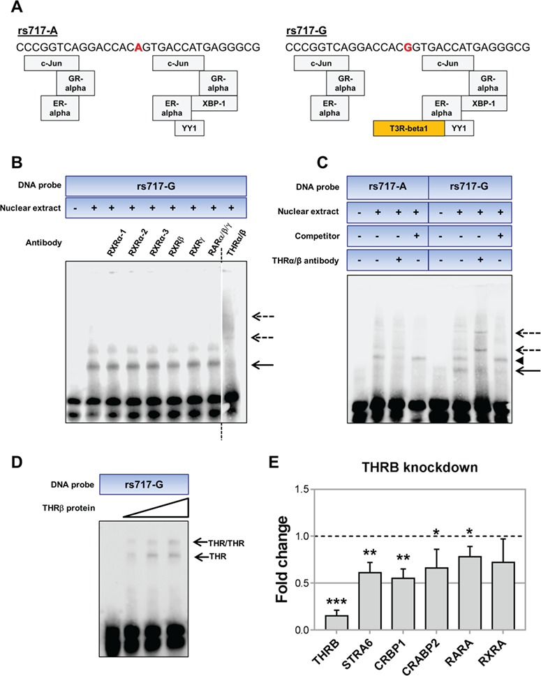
LOXL1 (lysyl oxidase-like 1) has been identified as the major effect locus in pseudoexfoliation (PEX) syndrome, a fibrotic disorder of the extracellular matrix and frequent cause of chronic open-angle glaucoma. However, all known PEX-associated common variants show allele effect reversal in populations of different ancestry, casting doubt on their biological significance. Based on extensive LOXL1 deep sequencing, we report here the identification of a common non-coding sequence variant, rs7173049A>G, located downstream of LOXL1, consistently associated with a decrease in PEX risk (odds ratio, OR = 0.63; P = 6.33 × 10⁻³¹) in nine different ethnic populations. We provide experimental evidence for a functional enhancer-like regulatory activity of the genomic region surrounding rs7173049 influencing expression levels of ISLR2 (immunoglobulin superfamily containing leucine-rich repeat protein 2) and STRA6 [stimulated by retinoic acid (RA) receptor 6], apparently mediated by allele-specific binding of the transcription factor thyroid hormone receptor beta. We further show that the protective rs7173049-G allele correlates with increased tissue expression levels of ISLR2 and STRA6 and that both genes are significantly downregulated in tissues of PEX patients together with other key components of the STRA6 receptor-driven RA signaling pathway. siRNA-mediated downregulation of RA signaling induces upregulation of LOXL1 and PEX-associated matrix genes in PEX-relevant cell types. These data indicate that dysregulation of STRA6 and impaired retinoid metabolism are involved in the pathophysiology of PEX syndrome and that the variant rs7173049-G, which represents the first common variant at the broad LOXL1 locus without allele effect reversal, mediates a protective effect through upregulation of STRA6 in ocular tissues.


Figures

Figure 1
Figure 1
Forest plots for the association between rs7173049 and PEX syndrome in case–control cohorts from nine countries. (A) The effect of the rs7173049 G allele shows consistent odds ratios < 1 and is associated with decreased risk of PEX in all populations. (B) After adjustment for rs3825942, residual association of rs7173049 persists in the majority of cohorts. The horizontal lines denote the 95% confidence intervals of the odds ratio for each collection.
Figure 2
Figure 2
Location of SNP rs7173049 in an active regulatory genomic region. (A) Genomic region surrounding rs7173049 located downstream of LOXL1 viewed in UCSC genome browser (http://genome.ucsc.edu: GRCh37/hg19). ENCODE Regulation Tracks specify layered histone marks for H3K27Ac, DNase hypersensitivity regions and transcription factor binding sites, which indicate active regulatory elements flanking the SNP of interest, rs7173049. (B) Dual luciferase reporter assays demonstrating significantly increased regulatory activity for pGL4.23 [luc2/minP] reporter constructs containing a 200 bp fragment surrounding rs7173049 with either allele A (rs717-A) or G (rs717-G) upstream of a gene-unspecific minimal promoter compared to pGL4.23 [luc2/minP] empty vector without the rs7173049 insert in hTCFs (P < 0.01) and TM cells (hTMC; P < 0.05). Results are expressed as the ratio of Firefly luciferase to Renilla luciferase and analyzed in relation to the transcriptional activity of the pGL4.23 empty vector set at 1 (dashed line). (C) Relative luciferase activity of pGL4.10-LOXL1 reporter constructs containing a 200 bp fragment surrounding rs7173049 with either allele A (rs717-A) or G (rs717-G) upstream of the specific LOXL1 core promoter was not increased over basal pGL4.10-LOXL1 promoter activity without the rs7173049 insert in hTCF and hTMC. Results are expressed as the ratio of Firefly luciferase to Renilla luciferase and analyzed in relation to the transcriptional activity of the pGL4.10-LOXL1 basal vector set at 1 (dashed line). (D) EMSAs using 31 bp biotinylated DNA probes containing rs7173049 A (rs717-A) or G (rs717-G) allele and nuclear extracts from hTCF and hTMC showing specific DNA–protein complexes (arrows). Following titration with increasing concentrations of nuclear proteins (left), shifted bands could be completely inhibited by unlabeled oligonucleotides proving specificity (right). Quantitative analysis of the shifted bands relative to the unshifted bands revealed allele-specific differences with significantly stronger binding of the G allele compared to the A allele set at 100%. Data represent mean values ± standard deviation (SD) of at least three independent experiments (*P < 0.05; **P < 0.01; ***P < 0.001).
Figure 3
Figure 3
Transcriptional regulation of LOXL1, STRA6 and ISLR2 by genomic region containing rs7173049. (A) Schematic diagram showing the genomic region 1 Mb up- and downstream of rs7173049 (chr15:73244610-75244610) containing 31 genes. (B) Quantitative real-time PCR assays of all 31 neighboring genes showing significant downregulation of LOXL1 as well as significant upregulation of STRA6 and ISLR2 in HEK293T cells after deletion of a 200 bp region flanking rs7173049 using CRISPR/Cas9 technology. Data represent mean values of eight edited (Deletion) and six non-edited (Control) clones; relative expression levels were normalized to GAPDH and are represented as means ± SD (*P < 0.05; **P < 0.01). (C) Immunofluorescence staining of HEK293T cells confirming reduced protein expression of LOXL1 as well as increased protein expression of STRA6 and ISLR2 in genome-edited cells compared to non-edited control cells. Nuclei are counterstained with DAPI (blue); scale bars are equal to 25 μm.
Figure 4
Figure 4
Correlation of rs7173049 genotype and tissue expression levels of STRA6 and ISLR2. (A) Genotype-correlated mRNA expression levels of STRA6 and ISLR2 in human iris tissue samples homozygous for allele A (n = 33) or allele G (n = 5) or heterozygous for both alleles (n = 28) at rs7173049 using real-time PCR technology. Expression levels of STRA6 and ISLR2 are significantly increased in specimens homozygous for the protective allele G compared to samples with A/G and A/A genotype. (B) Expression of STRA6 and ISLR2 mRNA in ocular tissues (iris and retina) derived from normal human donors (control, n = 42) and donors with PEX syndrome (n = 24) using real-time PCR technology. Expression levels were significantly reduced in PEX specimens compared to control specimens. The relative expression levels were normalized relative to GAPDH and are represented as mean values ± SD (*P < 0.05; **P < 0.01; ***P < 0.001). (C) Expression of STRA6 protein in ocular tissues (retina, iris and ciliary body) of normal human donor eyes and donor eyes with PEX syndrome, as determined by immunofluorescence labeling. In normal eye tissues (control), STRA6 immunopositivity (green fluorescence) is observed in the RPE overlying the choroid (CH), retinal BVs (asterisk) within the RGC layer, iridal BVs (asterisk) in the iris ST and the inner layer of the CE adjacent to the ciliary ST. Expression levels are reduced in tissues of PEX eyes, and reduced staining intensities are often associated with LOXL1-positive PEX material accumulations (PEX, red immunofluorescence) in iris blood vessel walls and on the surface of the CE. (D) Expression of ISLR2 protein in ocular tissues (retina, iris and TM) of normal human donor eyes and donor eyes with PEX syndrome, as determined by immunofluorescence labeling. In normal control tissues, ISLR2 immunopositivity (green fluorescence) is observed in the retinal NFL, single cells (arrow) of the retinal ganglion cell layer (RGC) adjacent to BVs (asterisk), endothelial cells of BVs (asterisks) in the iris ST, and endothelial cells of the TM and SC. Markedly reduced staining intensities were seen in tissues of PEX eyes (INL means inner nuclear layer; ONL, outer nuclear layer; DAPI nuclear counterstain in blue).
Figure 4
Figure 4
Continued
Figure 5
Figure 5
Diminished RA signaling in PEX tissues. (A) Expression of CRBP1, CRABP2, RARA and RXRA mRNA in iris (n = 42) and ciliary body specimens (n = 22) from normal human donors (control) and donors with PEX syndrome (n = 24) using real-time PCR technology. Expression levels were significantly reduced in PEX specimens compared to control specimens. The relative expression levels were normalized relative to GAPDH and are represented as mean values ± SD. (B) Western blot analysis of CRBP1 protein expression in iris and ciliary body tissue from normal donors (control, n = 4) and patients with PEX syndrome (n = 4). Protein expression is normalized to the house-keeping gene ß-actin and is expressed as percent of the expression in controls. (C) Western blot analysis of RBP4 protein in serum and aqueous humor samples from cataract patients (control, n = 4) and patients with PEX syndrome (n = 4). RBP4 protein is normalized to total protein content and is expressed as percent of controls (data represent mean values ± SD of eight samples; *P < 0.05; **P < 0.01 ***P < 0.001). (D) Real-time PCR analysis of RXRA, LOXL1, ELN, FBN1 and TGFB1 mRNA in hTCFs (n = 4) and TM cells (hTMC, n = 4) transfected with RXRA-specific siRNA or scrambled control siRNA; expression levels were normalized relative to GAPDH and expressed relative to mock-transfected controls (dashed line; *P < 0.05; **P < 0.01; ***P < 0.001). (E) Real-time PCR analysis of RXRA, LOXL1, ELN, FBN1 and TGFB1 mRNA in hTCF and hTMC without and with stimulation by 2.5 μm RA for 48 h; expression levels were normalized relative to GAPDH and expressed relative to unstimulated controls (dashed line; *P < 0.05; **P < 0.001; ***P < 0.0001).
Figure 6
Figure 6
Allele-specific transcription factor binding at the genomic region surrounding rs7173049.(A)Transcription factor binding sites overlapping SNP rs7173049 (marked in red) regions (±15 bp) as predicted by the PROMO bioinformatics program. Differential binding to sequences containing the protective G allele (rs717-G) compared to sequences containing the A allele (rs717-A) was only predicted for T3R-beta1 (THRβ1). Each gray/orange box indicates one transcription factor: GR-alpha, ER-alpha, XBP-1 and YY1. (B) Supershift assays with 31 bp biotinylated DNA probes containing rs7173049 G allele (rs717-G), nuclear extracts from hTCFs and specific antibodies against candidate transcription factors RXRα-1,2,3, beta (RXRβ) and gamma (RXRγ), RARα/β/γ and thyroid hormone receptor alpha/beta (THRα/β) showing disruption of the shifted DNA–protein complex (solid arrow) and formation of supershifted bands (dotted arrows) with the THRα/βantibody. Dotted black line indicates a cut and junction of two parts of the same membrane. (C) Supershift assays with THRα/β antibody attenuated the shifted DNA–protein complex (solid arrow) and produced distinct supershifted bands (dotted arrows) with DNA probes containing the protective allele G of rs7173049 (rs717-G). No supershifted bands were detected with probes containing the A allele (rs717-A). Bands which were not competitively inhibited by unlabeled oligonucleotides (arrowhead) were considered unspecific. (D) Increasing amounts of recombinant THRβ protein were used in positive control experiments to show a specific interaction with the DNA probe containing the protective G allele of rs7173049 (rs717-G). DNA–protein complexes (arrows) presumably represent thyroid hormone receptor (THR) monomers and homodimers. (E) Real-time PCR analysis of THRB, CRBP1, CRABP2, RARA and RXRA mRNA in hTCFs (n = 4) transfected with THRB-specific siRNA or scrambled control siRNA; expression levels were normalized relative to GAPDH and expressed relative to mock-transfected controls (dashed line; (*P < 0.05; **P < 0.01 ***P < 0.001).

Similar articles

Cited by

References

    1. Konstas A.G.P. and Ringvold A. (2018) Epidemiology of exfoliation syndrome. J. Glaucoma, 27, S4–S11. - PubMed
    1. Ritch R. and Schlötzer-Schrehardt U. (2001) Exfoliation syndrome. Surv. Ophthalmol., 45, 265–315. - PubMed
    1. Schlötzer-Schrehardt U. and Naumann G.O. (2006) Ocular and systemic pseudoexfoliation syndrome. Am. J. Ophthalmol., 141, 921–937. - PubMed
    1. Ozaki M. (2018) Mechanisms of glaucoma in exfoliation syndrome. J. Glaucoma, 27, S83–S86. - PubMed
    1. Ritch R. (1994) Exfoliation syndrome—the most common identifiable cause of open-angle glaucoma. J. Glaucoma, 3, 176–177. - PubMed

Publication types