. 2019 Apr 3;20(7):1647.
doi: 10.3390/ijms20071647.

AICAR Induces Apoptosis and Inhibits Migration and Invasion in Prostate Cancer Cells Through an AMPK/mTOR-Dependent Pathway

Affiliations

AICAR Induces Apoptosis and Inhibits Migration and Invasion in Prostate Cancer Cells Through an AMPK/mTOR-Dependent Pathway

Chia-Cheng Su et al. Int J Mol Sci. .

Abstract

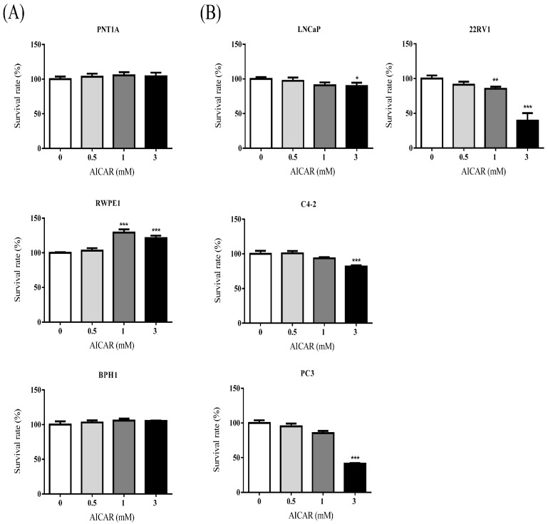
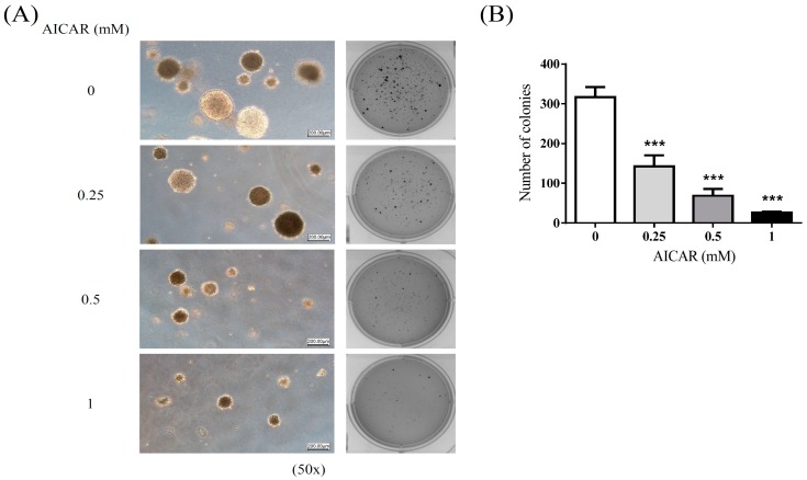
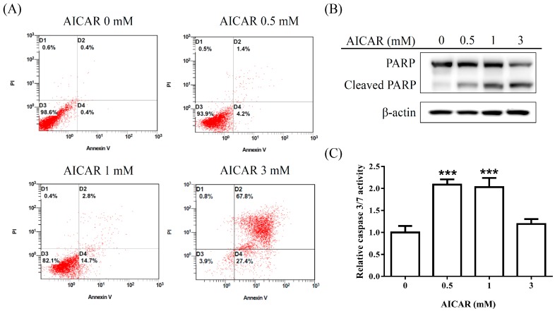
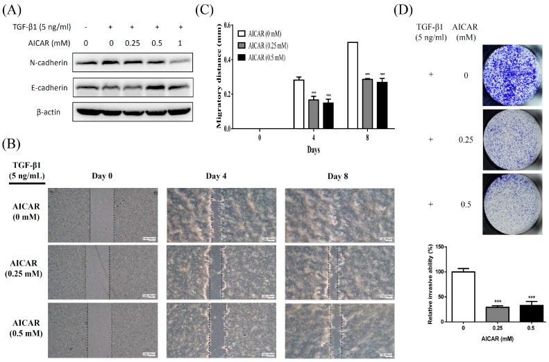
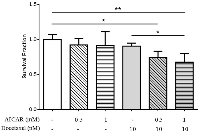
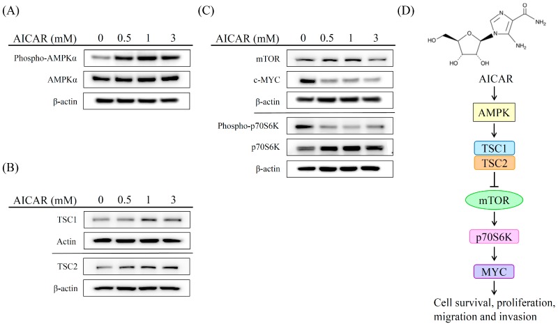
Current clinical challenges of prostate cancer management are to restrict tumor growth and prohibit metastasis. AICAR (5-aminoimidazole-4-carbox-amide-1-β-d-ribofuranoside), an AMP-activated protein kinase (AMPK) agonist, has demonstrated antitumor activities for several types of cancers. However, the activity of AICAR on the cell growth and metastasis of prostate cancer has not been extensively studied. Herein we examine the effects of AICAR on the cell growth and metastasis of prostate cancer cells. Cell growth was performed by MTT assay and soft agar assay; cell apoptosis was examined by Annexin V/propidium iodide (PI) staining and poly ADP ribose polymerase (PARP) cleavage western blot, while cell migration and invasion were evaluated by wound-healing assay and transwell assay respectively. Epithelial-mesenchymal transition (EMT)-related protein expression and AMPK/mTOR-dependent signaling axis were analyzed by western blot. In addition, we also tested the effect of AICAR on the chemosensitivity to docetaxel using MTT assay. Our results indicated that AICAR inhibits cell growth in prostate cancer cells, but not in non-cancerous prostate cells. In addition, our results demonstrated that AICAR induces apoptosis, attenuates transforming growth factor (TGF)-β-induced cell migration, invasion and EMT-related protein expression, and enhances the chemosensitivity to docetaxel in prostate cancer cells through regulating the AMPK/mTOR-dependent pathway. These findings support AICAR as a potential therapeutic agent for the treatment of prostate cancer.

Keywords: AICAR; AMPK; chemosensitivity; metastasis; prostate cancer.


Conflict of interest statement

The authors declare no conflict of interest.

Figures

Figure 1
Figure 1
The effect of AICAR (5-aminoimidazole-4-carbox-amide-1-β-d-ribofuranoside) on the growth of non-cancerous or cancerous prostate cells. (A) non-cancerous or (B) cancerous prostate cells were treated with different concentrations of AICAR for 24 h. The cell viability was measured by the MTT assay. Data are represented as means ± SD of triplicate values and statistical significance was determined using the Student’s t-test (* p < 0.05; ** p < 0.01; *** p < 0.001).
Figure 2
Figure 2
The effect of AICAR on growth under anchorage-independent conditions of prostate cancer cells. 22Rv1 cells were treated with different concentrations of AICAR, and then grown in soft agar, an anchorage-independent condition, for 3 weeks. (A) Colonies were stained with crystal violet and captured using the Bio-Rad ChemiDoc XRS+ system (Hercules, CA, USA). (B) Data are quantified and represented as means ± SD of triplicate values and statistical significance was determined using the Student’s t-test (*** p < 0.001).
Figure 3
Figure 3
Effect of AICAR on the apoptosis in 22Rv1 prostate cancer cells. Cells were incubated with different concentrations of AICAR for 24 h. (A) Cells were collected, stained with Annexin V and propidium iodide (PI), and analyzed by flow cytometry. Data are representative of at least three independent experiments with similar results. (B) The expression of Poly(ADP-ribose) polymerase (PARP) was determined by western blot. Actin was used as a loading control in western blot. (C) Cellular caspase 3/7 activities were analyzed with caspase-glo assay kit. Data are represented as means ± SD of triplicate values and statistical significance was determined using the Student’s t-test (*** p < 0.001).
Figure 4
Figure 4
Effect of AICAR on transforming growth factor-β (TGF)-β-induced epithelial to mesenchymal transition (EMT), migration, and invasion in 22Rv1 prostate cancer cells. (A) Cells were treated with 5 ng/mL TGF-β1 and different concentrations of AICAR for 72 h. The expression of N-cadherin and E-cadherin was analyzed by western blot. Actin was used as a loading control in western blot. The western blotting results are representative of results obtained in three separate experiments. (B) Cells were seeded in SPLScarTM Block overnight, the block was then removed, and cells were treated with 5 ng/mL TGF-β1 and different concentrations of AICAR. Cell migration was monitored under a phase-contrast microscope. Data are representative of at least three independent experiments with similar results. (C) The migratory distance was calculated. (D) Cells were treated with 5 ng/mL TGF-β1 and different concentrations of AICAR, then seeded into Matrigel transwell inserts for 5 days. The invaded cells were quantified using crystal violet staining. Data are represented as means ± SD of triplicate values and statistical significance was determined using the Student’s t-test (* p < 0.05; ** p < 0. 01; *** p < 0.001).
Figure 5
Figure 5
Effect of AICAR on the sensitivity of 22Rv1 prostate cancer cells to docetaxel treatment. Cells were treated with different concentrations of AICAR in the presence or absence of docetaxel for 24 h. The cell viability was measured by the MTT assay. Data are represented as means ± SD of triplicate values and statistical significance was determined using the Student’s t-test (* p < 0.05; ** p < 0.01).
Figure 6
Figure 6
Effect of AICAR on the AMPK/mTOR-dependent pathway in 22Rv1 prostate cancer cells. Cells were treated with different concentrations of AICAR for 2 h. The expression of (A) phospho-AMPK, AMPK, (B) TSC1 and TSC2 was examined by western blot. Cells were treated with different concentrations of AICAR for 6 h. (C) The expression of mTOR, cMYC, phosphor-p70S6K and p70S6K was determined by western blot. Actin was used as a loading control in western blot. The western blotting results are representative of results obtained in three separate experiments. (D) Proposed mechanism of the anticancer effects induced by AICAR in 22Rv1 prostate cancer cells.

References

    1. Siegel R.L., Miller K.D., Jemal A. Cancer statistics, 2018. CA Cancer J. Clin. 2018;68:7–30. doi: 10.3322/caac.21442. - DOI - PubMed
    1. Litwin M.S., Tan H.J. The diagnosis and treatment of prostate cancer: A review. JAMA. 2017;317:2532–2542. doi: 10.1001/jama.2017.7248. - DOI - PubMed
    1. Marshall S., Taneja S. Focal therapy for prostate cancer: The current status. Prostate Int. 2015;3:35–41. doi: 10.1016/j.prnil.2015.03.007. - DOI - PMC - PubMed
    1. Asmane I., Ceraline J., Duclos B., Rob L., Litique V., Barthelemy P., Bergerat J.P., Dufour P., Kurtz J.E. New strategies for medical management of castration-resistant prostate cancer. Oncology. 2011;80:1–11. doi: 10.1159/000323495. - DOI - PubMed
    1. Petrylak D.P., Tangen C.M., Hussain M.H., Lara P.N., Jr., Jones J.A., Taplin M.E., Burch P.A., Berry D., Moinpour C., Kohli M., et al. Docetaxel and estramustine compared with mitoxantrone and prednisone for advanced refractory prostate cancer. N. Engl. J. Med. 2004;351:1513–1520. doi: 10.1056/NEJMoa041318. - DOI - PubMed

MeSH terms