Protective Effect of Cashew Gum (Anacardium occidentale L.) on 5-Fluorouracil-Induced Intestinal Mucositis
- PMID: 30987265
- PMCID: PMC6630449
- DOI: 10.3390/ph12020051
Protective Effect of Cashew Gum (Anacardium occidentale L.) on 5-Fluorouracil-Induced Intestinal Mucositis
Abstract
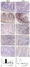
Intestinal mucositis is a common complication associated with 5-fluorouracil (5-FU), a chemotherapeutic agent used for cancer treatment. Cashew gum (CG) has been reported as a potent anti-inflammatory agent. In the present study, we aimed to evaluate the effect of CG extracted from the exudate of Anacardium occidentale L. on experimental intestinal mucositis induced by 5-FU. Swiss mice were randomly divided into seven groups: Saline, 5-FU, CG 30, CG 60, CG 90, Celecoxib (CLX), and CLX + CG 90 groups. The weight of mice was measured daily. After treatment, the animals were euthanized and segments of the small intestine were collected to evaluate histopathological alterations (morphometric analysis), levels of malondialdehyde (MDA), myeloperoxidase (MPO), and glutathione (GSH), and immunohistochemical analysis of interleukin 1 beta (IL-1β) and cyclooxygenase-2 (COX-2). 5-FU induced intense weight loss and reduction in villus height compared to the saline group. CG 90 prevented 5-FU-induced histopathological changes and decreased oxidative stress through decrease of MDA levels and increase of GSH concentration. CG attenuated inflammatory process by decreasing MPO activity, intestinal mastocytosis, and COX-2 expression. Our findings suggest that CG at a concentration of 90 mg/kg reverses the effects of 5-FU-induced intestinal mucositis.
Keywords: 5-fluorouracil; heteropolysaccharide; inflammation; intestinal mucositis.
Conflict of interest statement
The authors declare no conflict of interest.
Figures


Similar articles
-
Troxerutin Prevents 5-Fluorouracil Induced Morphological Changes in the Intestinal Mucosa: Role of Cyclooxygenase-2 Pathway.Pharmaceuticals (Basel). 2020 Jan 8;13(1):10. doi: 10.3390/ph13010010. Pharmaceuticals (Basel). 2020. PMID: 31936203 Free PMC article.
-
Role of Rutin in 5-Fluorouracil-Induced Intestinal Mucositis: Prevention of Histological Damage and Reduction of Inflammation and Oxidative Stress.Molecules. 2020 Jun 17;25(12):2786. doi: 10.3390/molecules25122786. Molecules. 2020. PMID: 32560278 Free PMC article.
-
Antidiarrheal activity of cashew GUM, a complex heteropolysaccharide extracted from exudate of Anacardium occidentale L. in rodents.J Ethnopharmacol. 2015 Nov 4;174:299-307. doi: 10.1016/j.jep.2015.08.020. Epub 2015 Aug 20. J Ethnopharmacol. 2015. PMID: 26297843
-
Cashew gum as future multipurpose biomacromolecules.Carbohydr Polym. 2025 Jan 1;347:122749. doi: 10.1016/j.carbpol.2024.122749. Epub 2024 Sep 17. Carbohydr Polym. 2025. PMID: 39486978 Review.
-
Cashew Gum: A Review of Brazilian Patents and Pharmaceutical Applications with a Special Focus on Nanoparticles.Micromachines (Basel). 2022 Jul 18;13(7):1137. doi: 10.3390/mi13071137. Micromachines (Basel). 2022. PMID: 35888956 Free PMC article. Review.
Cited by
-
Essential Oil-Containing Polysaccharide-Based Edible Films and Coatings for Food Security Applications.Polymers (Basel). 2021 Feb 14;13(4):575. doi: 10.3390/polym13040575. Polymers (Basel). 2021. PMID: 33672974 Free PMC article. Review.
-
Troxerutin Prevents 5-Fluorouracil Induced Morphological Changes in the Intestinal Mucosa: Role of Cyclooxygenase-2 Pathway.Pharmaceuticals (Basel). 2020 Jan 8;13(1):10. doi: 10.3390/ph13010010. Pharmaceuticals (Basel). 2020. PMID: 31936203 Free PMC article.
-
Losartan improves intestinal mucositis induced by 5-fluorouracil in mice.Sci Rep. 2021 Dec 1;11(1):23241. doi: 10.1038/s41598-021-01969-x. Sci Rep. 2021. PMID: 34853351 Free PMC article.
-
Special Issue "Anticancer Drugs".Pharmaceuticals (Basel). 2019 Sep 16;12(3):134. doi: 10.3390/ph12030134. Pharmaceuticals (Basel). 2019. PMID: 31527393 Free PMC article.
-
Protective Effects of Oxyberberine in 5-Fluorouracil-Induced Intestinal Mucositis in the Mice Model.Evid Based Complement Alternat Med. 2022 May 30;2022:1238358. doi: 10.1155/2022/1238358. eCollection 2022. Evid Based Complement Alternat Med. 2022. PMID: 35677366 Free PMC article.
References
-
- Kumar V., Abbas A.K., Aster J.C. Robbins Basic Pathology E-Book. 9th ed. Elsevier Health Sciences; Rio de Janeiro, Brazil: 2013.
-
- Ferreira J.E.V., de Figueiredo A.F., Barbosa J.P., Pinheiro J.C. Chemometrics in Practical Applications. IntechOpen; London, UK: 2012. Chemometric study on molecules with anticancer properties; pp. 185–186.
-
- Organization W.H. Cancer. [(accessed on 17 August 2018)]; Available online: http://www.who.int/cancer/en/
-
- Chang C.-T., Ho T.-Y., Lin H., Liang J.-A., Huang H.-C., Li C.-C., Lo H.-Y., Wu S.-L., Huang Y.-F., Hsiang C.-Y. 5-Fluorouracil induced intestinal mucositis via nuclear factor-κB activation by transcriptomic analysis and in vivo bioluminescence imaging. PLoS ONE. 2012;7:e31808. doi: 10.1371/journal.pone.0031808. - DOI - PMC - PubMed
LinkOut - more resources
Full Text Sources
Research Materials
Miscellaneous

