. 2019 Apr 15;9(1):6091.
doi: 10.1038/s41598-019-42572-5.

Genome analysis of Paenibacillus polymyxa A18 gives insights into the features associated with its adaptation to the termite gut environment

Affiliations

Genome analysis of Paenibacillus polymyxa A18 gives insights into the features associated with its adaptation to the termite gut environment

Nandita Pasari et al. Sci Rep. .

Abstract

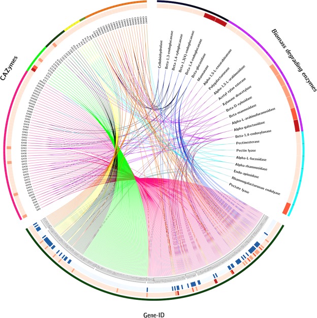
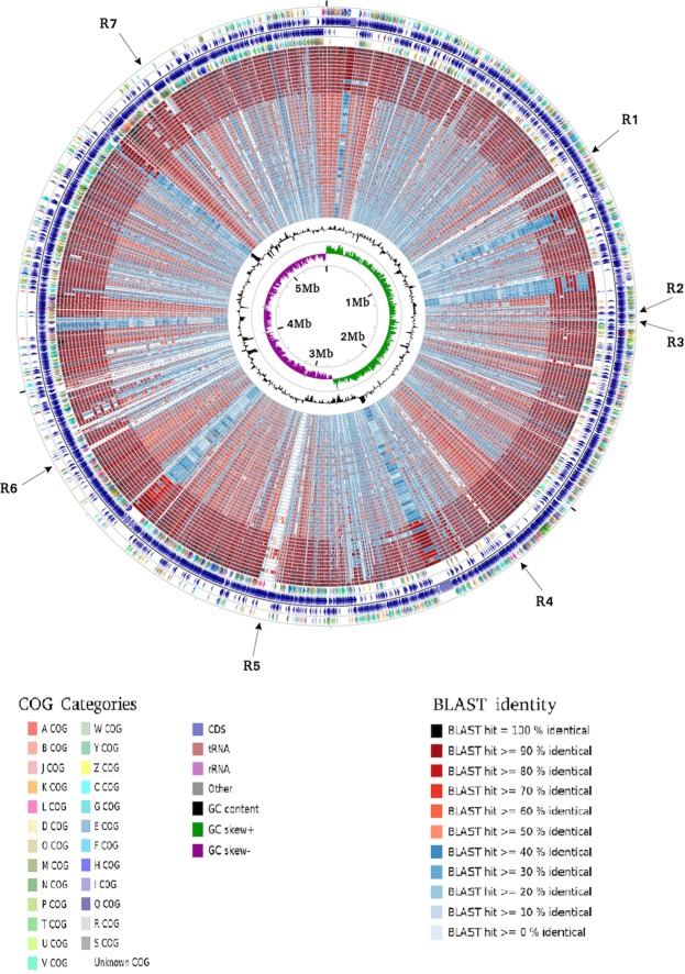
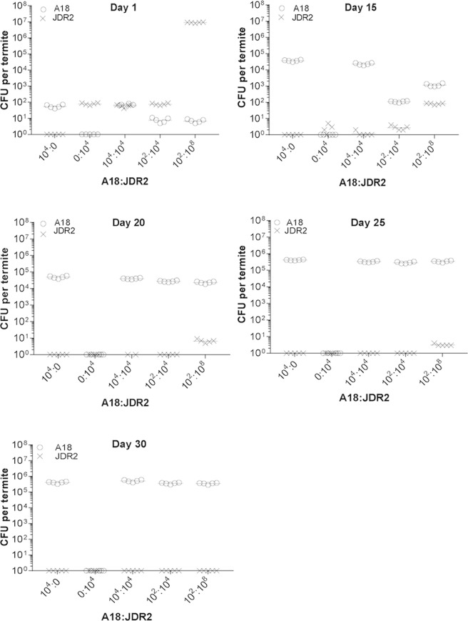
Paenibacillus polymyxa A18 was isolated from termite gut and was identified as a potential cellulase and hemicellulase producer in our previous study. Considering that members belonging to genus Paenibacillus are mostly free-living in soil, we investigated here the essential genetic features that helped P. polymyxa A18 to survive in gut environment. Genome sequencing and analysis identified 4608 coding sequences along with several elements of horizontal gene transfer, insertion sequences, transposases and integrated phages, which add to its genetic diversity. Many genes coding for carbohydrate-active enzymes, including the enzymes responsible for woody biomass hydrolysis in termite gut, were identified in P. polymyxa A18 genome. Further, a series of proteins conferring resistance to 11 antibiotics and responsible for production of 4 antibiotics were also found to be encoded, indicating selective advantage for growth and colonization in the gut environment. To further identify genomic regions unique to this strain, a BLAST-based comparative analysis with the sequenced genomes of 47 members belonging to genus Paenibacillus was carried out. Unique regions coding for nucleic acid modifying enzymes like CRISPR/Cas and Type I Restriction-Modification enzymes were identified in P. polymyxa A18 genome suggesting the presence of defense mechanism to combat viral infections in the gut. In addition, genes responsible for the formation of biofilms, such as Type IV pili and adhesins, which might be assisting P. polymyxa A18 in colonizing the gut were also identified in its genome. In situ colonization experiment further confirmed the ability of P. polymyxa A18 to colonize the gut of termite.


Conflict of interest statement

The authors declare no competing interests.

Figures

Figure 1
Figure 1
Circular map of Paenibacillus polymyxa A18 genome. From outer circle to inner circle, representation is as follows: 1. Position in megabases (black); 2. Forward strand CDSs (turquoise blue); 3. Reverse strand CDSs (turquoise blue); 4. Horizontal gene transfer (HGT) regions (red); 5. Insertion sequences (IS) (blue); 6. Phage sequences (grey); 7. tRNAs (pink); 8. GC plot (mustard and blue colour correspond to higher and lower than average GC content, respectively); 9. GC skew (mustard and blue colour correspond to higher and lower than average GC-skew, respectively).
Figure 2
Figure 2
Phylogenetic analyses of the members of the genus Paenibacillus. Maximum-likelihood phylogenetic tree computed using the core genes of completely sequenced Paenibacillus sp. generated with 1000 bootstrap replications. Scale bar indicates the average number of substitutions per site.
Figure 3
Figure 3
Comparative genome analyses of the members of the genus Paenibacillus. P. polymyxa A18 genome comparison with completely sequenced members of the genus Paenibacillus. Each of the genome sequence assemblies (the GenBank accession numbers are listed in Supplementary Table S3 in the order of their presence in the concentric rings) were aligned against the P. polymyxa A18 genome. The innermost ring indicates the genomic position. The next two rings represent G + C content and GC skew. The remaining concentric rings indicate the presence or absence of BLASTN hits at that position, with each ring corresponding to the genome assemblies in the order mentioned in Supplementary Table S3. Positions covered by BLASTN alignments are indicated with a solid color; white gaps represent genomic regions not covered by the BLASTN alignments. The graphical view of the alignments was rendered using BLAST Ring Image Generator (BRIG).
Figure 4
Figure 4
Carbohydrate-active enzyme (CAZymes) encoding genes present in P. polymyxa A18 genome including biomass degrading enzymes are represented. From outer to inner circle – Circle 1 - (a) Different CAZymes families GH (pink), GT (light green), CE (dark green), PL (yellow), AA (moss green) and associated module CBM (orange); (b) Classification on the basis of biomass degrading abilities-cellulose degrading enzymes (dark blue), hemicellulose degrading enzymes (purple) and pectin degrading enzymes (cyan); (c) Gene Ids of all the CAZyme genes in P. polymyxa A18. Circle 2 - Heatmap of different enzyme types (represented in light to dark red based on its count in the genome). Circle 3 - CAZymes having a signal peptide (represented in dark blue).
Figure 5
Figure 5
Circular representation of P. polymyxa A18 CDSs and their alignment with CDSs of members of the genus Paenibacillus. From outer to inner circle: the 1st ring represents coding DNA sequences (CDS) of the plus strand of the P. polymyxa A18 chromosome under COG categories, the 2nd ring represents total CDS of the plus strand, the 3rd ring represents the total CDS of the minus strand, the 4th ring represents CDS of the minus strand under COG categories, and the 5th to 51st rings represent identity matches of CDS of the strains with P. polymyxa A18 as mentioned in the order in Supplementary Table S3. The innermost rings represent the average GC content of the plus and minus strands and the GC-skew, respectively. Regions relevant to gut colonization have been represented as R1 to R7. Details have been mentioned in Table 2.
Figure 6
Figure 6
Host specificity of P. polymyxa A18 as shown by colonization efficiency in termite. Gut bacterial load (CFU) was counted from day 1 after oral inoculation. The sample size of termite for each treatment was 5. Means of bacterial counts from 5 termites are shown as a straight line. The ratios of P. polymyxa A18 and Paenibacillus JDR-2 (represented as A18: JDR2) used in various experiments are shown in the X-axis.

References

    1. Chen, H. In Biotechnology of lignocellulose 25–71 (Springer, 2014).
    1. Ohkuma M. Termite symbiotic systems: efficient bio-recycling of lignocellulose. Applied microbiology and biotechnology. 2003;61:1–9. doi: 10.1007/s00253-002-1189-z. - DOI - PubMed
    1. Brune A, Dietrich C. The gut microbiota of termites: digesting the diversity in the light of ecology and evolution. Annual review of microbiology. 2015;69:145–166. doi: 10.1146/annurev-micro-092412-155715. - DOI - PubMed
    1. Warnecke F, et al. Metagenomic and functional analysis of hindgut microbiota of a wood-feeding higher termite. Nature. 2007;450:560–565. doi: 10.1038/nature06269. - DOI - PubMed
    1. Tokuda G, Watanabe H. Hidden cellulases in termites: revision of an old hypothesis. Biology Letters. 2007;3:336–339. doi: 10.1098/rsbl.2007.0073. - DOI - PMC - PubMed

Publication types