. 2019 Apr 15;9(1):6056.
doi: 10.1038/s41598-019-42403-7.

Birth and Resuscitation of (p)ppGpp Induced Antibiotic Tolerant Persister Cells

Affiliations

Birth and Resuscitation of (p)ppGpp Induced Antibiotic Tolerant Persister Cells

Mikkel Skjoldan Svenningsen et al. Sci Rep. .

Abstract

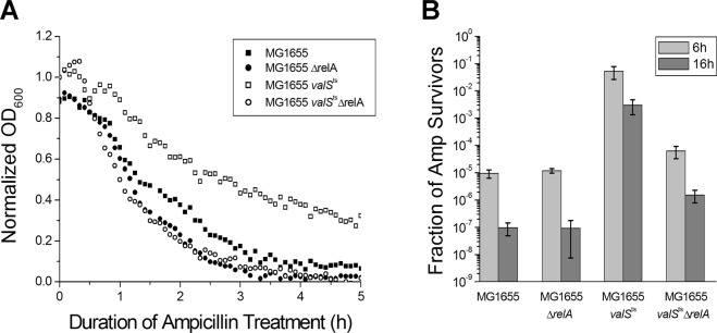
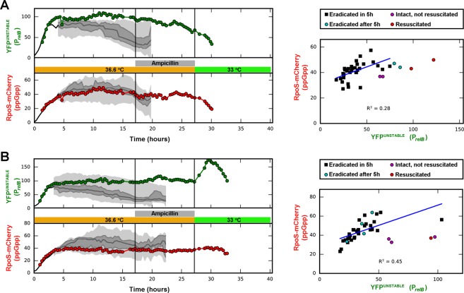
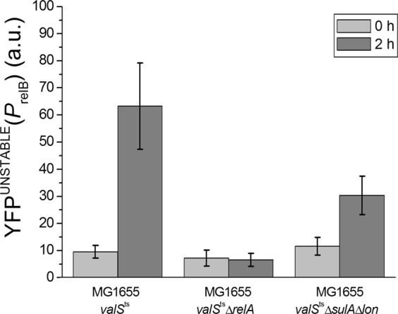
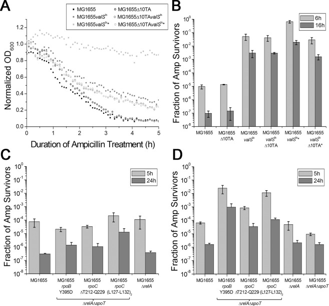
Transient antibiotic treatment typically eradicates most sensitive bacteria except a few survivors called persisters. The second messenger (p)ppGpp plays a key role in persister formation in Escherichia coli populations but the underlying mechanisms have remained elusive. In this study we induced (p)ppGpp synthesis by modulating tRNA charging and then directly observed the stochastic appearance, antibiotic tolerance, and resuscitation of persister cells using live microscopy. Different physiological parameters of persister cells as well as their regularly growing ancestors and sisters were continuously monitored using fluorescent reporters. Our results confirmed previous findings that high (p)ppGpp levels are critical for persister formation, but the phenomenon remained strikingly stochastic without any correlation between (p)ppGpp levels and antibiotic tolerance on the single-cell level. We could not confirm previous notions that persisters exhibit markedly low concentrations of intracellular ATP or were linked to post-transcriptional effects of (p)ppGpp through the activation of small genetic elements known as toxin-antitoxin (TA) modules. Instead, we suggest that persister cell formation under regular conditions is driven by the transcriptional response to increased (p)ppGpp levels.


Conflict of interest statement

The authors declare no competing interests.

Figures

Figure 1
Figure 1
RelA dependent persister cell formation induced by limitation of tRNA charging. Stationary phase MG1655valSts cells were diluted 1:1000 and grown under semi-permissive conditions (at 36.6 °C) until OD600 ~ 0.1 in a temperature-controlled microtiter plate reader, in supplemented M9 medium. MG1655, MG1655ΔrelA, and MG1655ΔrelAvalSts cells were used as controls. (A) Cells were treated with 150 μg/ml ampicillin, and the optical density of the cultures was recorded over time. The continuous decrease of OD600 over the 16 h incubation with ampicillin indicates that no resistant mutants appeared in the samples. (B) Colony forming units were determined before and at different time points during treatment; the ratios of survivors after 6 h and 16 h of treatment were plotted.
Figure 2
Figure 2
Single cell analysis of persister cell formation, ampicillin survival, and resuscitation in two micro-colonies (A,B). Left panels: MG1655valStsrpoS::mcherry cells carrying the plasmid borne relB promoter-yfpunstable fusion (pSEM4102) were grown in a temperature controlled microscope at semi-permissive temperature (36.6 °C). Micro-colonies that developed from single cells were treated with ampicillin. After ampicillin treatment, the antibiotic was removed and the temperature was lowered to 33 °C, allowing resuscitation of the antibiotic survivors. Fluorescence of relB promoter (YFPunstable) and the (p)ppGpp (RpoS-mCherry) reporters was recorded for all cells. Traces for two persister cells are shown in color. The traces end at first division after resuscitation, and the last data point represents the mean fluorescence of the two progeny cells after division. The median values of fluorescence are plotted along with the 25th, 50th, 75th and 100th percentiles of the non-persister cells of the colony. The percentiles are shown when there are 8 or more cells in the colony. Right panels: Distribution of fluorescence levels in the cells of the corresponding micro-colonies right before addition of ampicillin. The linear fit represents the correlation of the two markers in the non-persister population. More trajectories and corresponding pre-ampicillin distributions are shown in Fig. S2B. M9 medium supplemented by 3.2% glycerol and 17 amino acids (0.1 mg/mL) were used.
Figure 3
Figure 3
Changes in the levels of the relB promoter reporter (YFPunstable) upon limitation of tRNA charging in MG1655valSts, MG1655valStsΔsulAΔlon, and MG1655valStsΔrelA cells carrying the plasmid borne relB promoter-yfpunstable fusion (pSEM4102). Cells were grown in a temperature-controlled microscope on supplemented M9 agarose pads as described in Fig. 2. Images were taken at 0 time and after 2 hours growth at 36.6 °C. Similar results were obtained upon treatment with the seryl-tRNA synthetase inhibitor serine hydroxamate (SHX) at permissive temperature, indicating that they are not linked to the MG1655valSts strain or temperature stress but truly reflect the biology of limited tRNA charging (Fig. S3A).
Figure 4
Figure 4
Intracellular ATP concentration is not correlated with (p)ppGpp induced persistence. E. coli MG1655valSts cells carrying the pSEM4157 plasmid were grown in a temperature-controlled microscope as described for the experiment presented in Fig. 2. The dynamics of the PrelB promoter were followed by measuring the fluorescence of the mCherryunstable protein, transcribed from the PrelB promoter in plasmid pSEM4157. The intracellular ATP concentration was determined from the QUEEN-7µ fluorescence by calculating the ratio of the emitted light intensities at 520 nm using 405 and 490 nm excitation wavelengths. To follow the development of micro-colonies from single cells and appearance of persisters, QUEEN-7µ and PrelB-mCherry fluorescence levels were recorded every 20 minutes. Colonies were allowed to develop for 15 h and were then treated with ampicillin for 10 h. After removal of ampicillin, survivor cells were allowed to grow under permissive conditions (at 33 °C). Cells that were able to divide within 13 h were considered persisters. ATP levels and the level of the PrelB-mCherryunstable reporter were analyzed in colonies before ampicillin treatment (A–D). Red dots correspond to persister cells. ATP concentrations were calculated as described in Materials and Methods. The development of ATP and mCherryunstable levels during resuscitation of persisters is shown in Fig. S4.
Figure 5
Figure 5
Resuscitation of persisters is correlated with cessation of relBE transcription. MG1655valStsrpoS::mcherry cells carrying the pSEM4102 plasmid were grown in LB medium at 37 °C for about 6 hours (to mid-log phase) and treated with 100 µg/ml ampicillin for 3 hours, also at 37 °C. Survivor cells were concentrated by centrifugation and placed on a supplemented M9 agarose pad that contained β-lactamase to degrade any remaining ampicillin carried over from the growth medium. Changes in the cellular levels of YFPunstable and RpoS-mCherry were followed by fluorescence microscopy at 33 °C. Images were taken at the indicated time points. Combined mCherry (red) and YFP (green) channels are shown. Quantification of mCherry and YFPunstable fluorescence for 28 individual cells is shown in Fig. S5.
Figure 6
Figure 6
(A,B) Persister cell formation upon limitation of tRNA charging in the strain lacking the 10 mRNase toxins (Δ10TA). Stationary phase MG1655valSts cells were diluted 1:500 and grown at 36.6 °C until OD600 ~ 0.1 in a temperature-controlled microtiter plate reader. MG1655, MG1655Δ10TA, and MG1655valStsΔ10TA cells were used as controls. Cells were treated with 150 μg/ml ampicillin, and the optical density of the cultures was recorded over time (A). Colony forming units were counted before the treatment and 6 h and 16 h after treatment (B). Cells were treated with β-lactamase before plating on LB plates. Cultures marked with a star were shifted to room temperature for 5 minutes after 4 h of incubation and incubated for an additional ~2 h (until they reached OD600 ~ 0.1) before ampicillin treatment. (C,D) Antibiotic survival of MG1655 ΔrelA ΔspoT stringent mutants in supplemented M9 medium (C) or LB medium (D). Stationary phase cultures of the indicated strains were diluted 1:100 into fresh medium and directly challenged with 100 μg/ml ampicillin. Colony forming units were determined over time for 24 h, revealing biphasic killing. Bar diagrams display average and standard deviation of bacterial survival after 24 h of treatment. The growth of all strains in both media is shown in Fig. S6.

References

    1. Elowitz MB, Levine AJ, Siggia ED, Swain PS. Stochastic gene expression in a single cell. Science. 2002;297:1183–1186. doi: 10.1126/science.1070919. - DOI - PubMed
    1. Sneppen K, Krishna S, Semsey S. Simplified models of biological networks. Annu Rev Biophys. 2010;39:43–59. doi: 10.1146/annurev.biophys.093008.131241. - DOI - PubMed
    1. Rocha EP, Matic I, Taddei F. Over-representation of repeats in stress response genes: a strategy to increase versatility under stressful conditions? Nucleic Acids Res. 2002;30:1886–1894. doi: 10.1093/nar/30.9.1886. - DOI - PMC - PubMed
    1. Veening JW, Smits WK, Kuipers OP. Bistability, epigenetics, and bet-hedging in bacteria. Annu Rev Microbiol. 2008;62:193–210. doi: 10.1146/annurev.micro.62.081307.163002. - DOI - PubMed
    1. Hoyland-Kroghsbo NM, Maerkedahl RB, Svenningsen SL. A quorum-sensing-induced bacteriophage defense mechanism. MBio. 2013;4:e00362–00312. doi: 10.1128/mBio.00362-12. - DOI - PMC - PubMed

Publication types

MeSH terms