

HIGH TIBIAL OSTEOTOMY USING A LOCKING TITANIUM PLATE WITH OR WITHOUT AUTOGRAFTING
- PMID: 30988651
- PMCID: PMC6442715
- DOI: 10.1590/1413-785220192702164465
HIGH TIBIAL OSTEOTOMY USING A LOCKING TITANIUM PLATE WITH OR WITHOUT AUTOGRAFTING
Abstract
Objective: To postoperatively evaluate knee scores, radiological assessment results, deficit correction, patellar height change, bone healing time, and weight bearing time in patients undergoing high tibial osteotomy (HTO) with/without autologous iliac bone grafting.
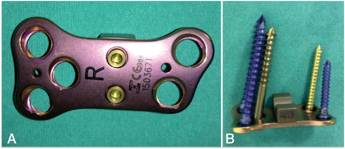
Methods: This retrospective examination of treated controls from a randomized controlled study included 63 knees of 58 patients aged 46-59 years who underwent HTO with locking open wedge osteotomy plates. The patients were divided into two groups: Group A, HTO with autologous iliac bone grafts (n = 31); and Group B, HTO without autologous iliac bone grafts (n = 32). Clinical and radiological data were evaluated prospectively at the preoperative consultation and again at 6, 9, and 12 weeks, 6 months, and 1 year after the surgery (and annually thereafter).
Results: There were no significant intergroup differences in the radiological assessment, deficit correction, patellar height change, bone-healing time, and weight-bearing time at any time after surgery. The knee scores changed positively in both groups (p < 0.001).
Conclusions: There was no difference in the results of patients undergoing HTO with open wedge osteotomy titanium locking plates with or without autografting, and comorbidities resulting from autografts were eliminated with the use of locking plates. Level of evidence III, Retrospective Study .
Objetivo: Avaliar escores de joelho, resultados da avaliação radiológica, correção de déficits, alteração da altura patelar, tempo de consolidação óssea e tempo para apoio de peso no pós-operatório em pacientes submetidos à osteotomia tibial alta (OTA) com ou sem enxerto autólogo de osso ilíaco.
Métodos: O exame retrospectivo de controles tratados em estudo randomizado e controlado foi realizado em 63 joelhos de 58 pacientes com idade entre 46 e 59 anos submetidos a OTA com placas bloqueadas de titânio em cunha aberta. Os pacientes foram divididos em dois grupos: Grupo A, OTA com enxerto de osso ilíaco autólogo (n = 31) e Grupo B, OTA sem enxerto autólogo de osso ilíaco (n = 32). Os dados clínicos e radiológicos foram avaliados prospectivamente na consulta pré-operatória e 6, 9 e 12 semanas e 6 meses e 1 ano após a cirurgia (e depois disso, anualmente).
Resultados: Não houve diferenças significativas quanto a avaliação radiológica, correção de déficit, mudança de altura da patela, tempo de cicatrização óssea e tempo para apoio de peso entre os dois grupos em nenhum momento após a cirurgia. Os escores de joelho mudaram positivamente em ambos os grupos (p < 0,001).
Conclusões: Não houve diferença nos resultados dos pacientes submetidos a OTA com placas bloqueadas de titânio em cunha aberta com e sem autoenxerto, e as comorbidades resultantes dos autoenxertos foram eliminadas com o uso de placas bloqueadas. Nível de Evidência III, Estudo Retrospectivo.
Keywords: Autograft; Bone plate; Correction; Healing; Osteotomy; Tibia.
Conflict of interest statement
All authors declare no potential conflict of interest related to this article.
Figures


References
-
- Gomes JLE, Ruthner RP, Marczyk LRS. Valgus tibial osteotomy with” wedge” plate of Puddu: technique presentation. Acta Ortop Bras. 2000;8(3):134–139.
-
- Jackson J. Osteotomy for osteoarthritis of the knee. J Bone Joint Surg Br. 1958;40(4):826–836.
-
- Coventry MB. Osteotomy of the upper portion of the tibia for degenerative arthritis of the knee. A preliminary report. 1965. J Bone Joint Surg Am. 1989;47(5):984–990. - PubMed
-
- Longino PD, Birmingham TB, Schultz WJ, Moyer RF, Giffin JR. Combined tibial tubercle osteotomy with medial opening wedge high tibial osteotomy minimizes changes in patellar height: a prospective cohort study with historical controls. Am J Sports Med. 2013;41(12):2849–2857. - PubMed
LinkOut - more resources
Full Text Sources