. 2019 Mar 25:15:30.
doi: 10.1186/s13007-019-0417-9. eCollection 2019.

Week-long imaging of cell divisions in the Arabidopsis root meristem

Affiliations

Week-long imaging of cell divisions in the Arabidopsis root meristem

Ramin Rahni et al. Plant Methods. .

Abstract

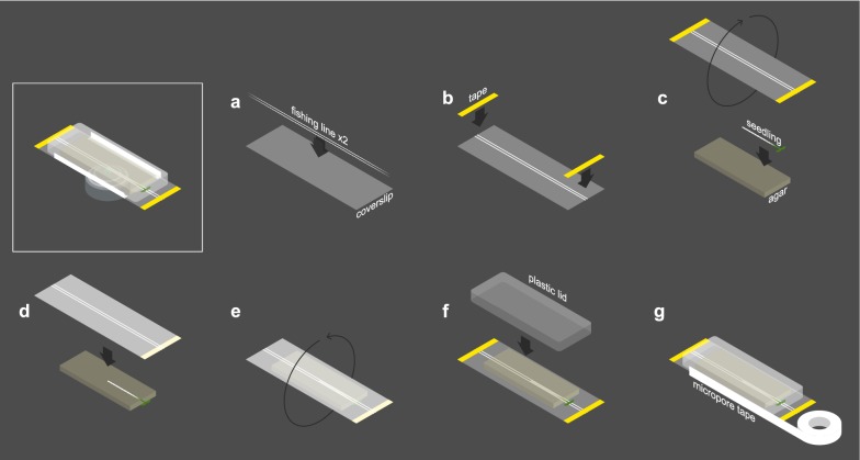
Background: Characterizing the behaviors of dynamic systems requires capturing them with high temporal and spatial resolution. Owing to its transparency and genetic tractability, the Arabidopsis thaliana root lends itself well to live imaging when combined with cell and tissue-specific fluorescent reporters. We developed a novel 4D imaging method that utilizes simple confocal microscopy and readily available components to track cell divisions in the root stem cell niche and surrounding region for up to 1 week.

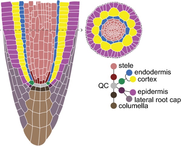
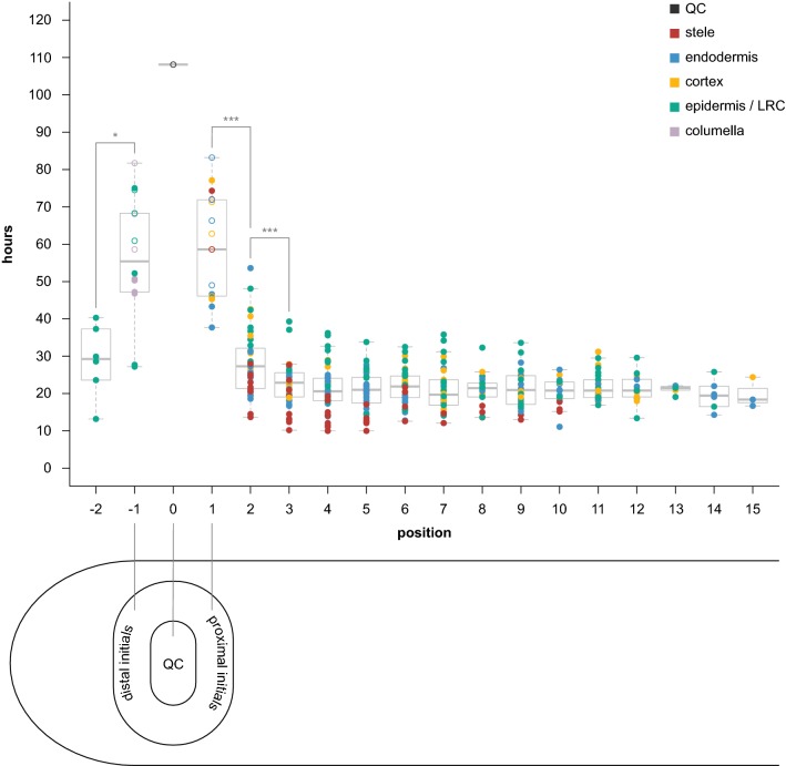
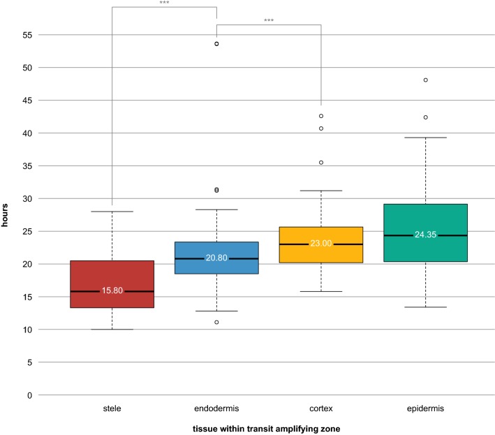
Results: Using this method, we performed a direct measurement of cell division intervals within and around the root stem cell niche. The results reveal a short, steep gradient of cell division rates in proximal stem cells, with progressively more rapid cell division rates from quiescent center (QC), to cells in direct contact with the QC (initials), to their immediate daughters, after which division rates appear to become more homogeneous.

Conclusions: These results provide a baseline to study how perturbations in signaling could affect cell division patterns in the root meristem. This new setup further allows us to finely analyze meristematic cell division rates that lead to patterning.

Keywords: Cell division; Confocal; Development; Live imaging; QC; Root; Stem cells; Time lapse.


Conflict of interest statement

The authors declare that they have no competing interests.

Figures

Fig. 1
Fig. 1
Schematic of a median cross section (left) of the Arabidopsis root apical meristem, showing the tissue-specific stem cells/initials surrounding the QC. Top-right shows a radial view of the root
Fig. 2
Fig. 2
Assembling the growth chamber. a Two strands of fishing line are placed against a full-length coverslip, approximately 1 mm apart. b The fishing line is affixed to the coverslip with thin strips of laboratory tape. c Seedling is laid against agar block while coverslip is rotated such that the fishing line side faces the seedling. d Coverslip is laid against seedling/agar, which is facing up to facilitate positioning the root inside the 1 mm gap between fishing line strands. The seedling is thus sandwiched between the agar and coverslip. e Seedling, agar, and coverslip combination are rotated (flipped over) such that the agar block is now on top of the coverslip. f A plastid lid is placed over the seedling and agar block to prevent them from drying out. g The points of contact between lid and coverslip are sealed with micropore tape to further ensure moisture retention and prevent contamination
Fig. 3
Fig. 3
Results of time-lapse experiments, showing hours between divisions by cellular position. Cells are color-coded by tissue type (legend in upper right-hand corner). Closed circles represent two “bookended” observed divisions for the same cell. Open circles denote a cell with only one observed division throughout the course of the film, with “hours” representing the time from that division until the beginning or end of the film (whichever happened to be longer). QC is denoted position 0. Proximal initials (stele, endodermis, cortex) are position 1. Distal initials (columella, epidermis/lateral root cap) are position − 1. Positions 2 and − 2 are the immediate daughters proximal and distal initials, respectively. Data represents divisions observed in four biological replicates (four separate films of four separate roots). Stars represent statistical significance (*p < 0.01, ***p < 0.0001)
Fig. 4
Fig. 4
Results of cell cycle duration within the Transit Amplifying zone (positions 2–15 in Fig. 3), sorted by tissue type. Data represents divisions observed in four biological replicates (four separate films of four separate roots). The value displayed on each box plot represents the median. Stars represent statistical significance (***p < 0.0001)

References

    1. Choe G, Lee J. Push–pull strategy in the regulation of postembryonic root development. Curr Opin Plant Biol [Internet] 2017;35:158–164. doi: 10.1016/j.pbi.2016.12.005. - DOI - PubMed
    1. Dolan L, Janmaat K, Willemsen V, Linstead P, Poethig S, Roberts K, et al. Cellular organisation of the Arabidopsis thaliana root. Development 1993;119:71–84. http://www.ncbi.nlm.nih.gov/pubmed/8275865. - PubMed
    1. Cruz-Ramírez A, Díaz-Triviño S, Wachsman G, Du Y, Arteága-Vázquez M, Zhang H, et al. A SCARECROW-RETINOBLASTOMA protein network controls protective quiescence in the Arabidopsis root stem cell organizer. PLoS Biol [Internet]. 2013;11:e1001724. http://www.pubmedcentral.nih.gov/articlerender.fcgi?artid=3841101&tool=p.... - PMC - PubMed
    1. Campilho A, Garcia B, Toorn HVD, Wijk HV, Campilho A, Scheres B. Time-lapse analysis of stem-cell divisions in the Arabidopsis thaliana root meristem. Plant J. 2006;48:619–627. doi: 10.1111/j.1365-313X.2006.02892.x. - DOI - PubMed
    1. Sena G, Frentz Z, Birnbaum KD, Leibler S. Quantitation of cellular dynamics in growing Arabidopsis roots with light sheet microscopy. PLoS One. 2011 [cited 2014 Jan 10];6:e21303. http://www.pubmedcentral.nih.gov/articlerender.fcgi?artid=3120859&tool=p.... - PMC - PubMed

LinkOut - more resources