. 2019 Mar 22:10:100203.
doi: 10.1016/j.bonr.2019.100203. eCollection 2019 Jun.

PI3K activation increases SDF-1 production and number of osteoclast precursors, and enhances SDF-1-mediated osteoclast precursor migration

Affiliations

PI3K activation increases SDF-1 production and number of osteoclast precursors, and enhances SDF-1-mediated osteoclast precursor migration

Naga Suresh Adapala et al. Bone Rep. .

Abstract

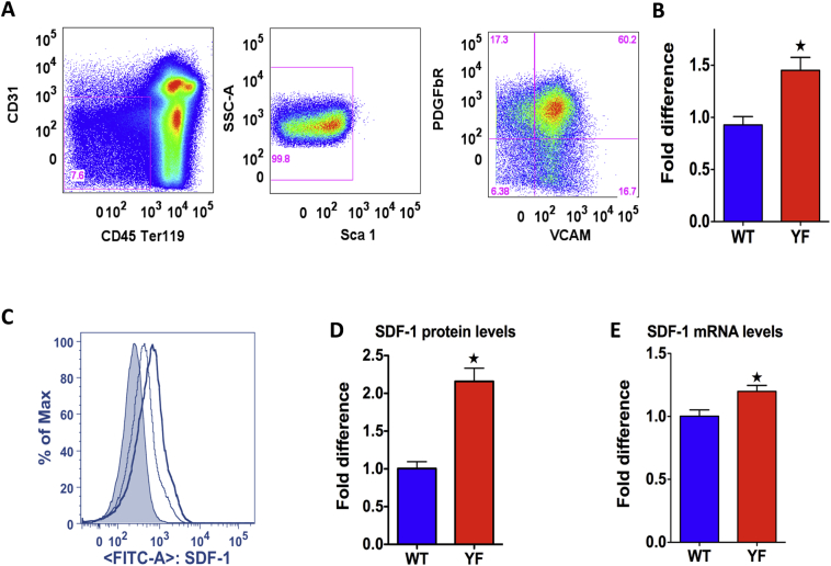
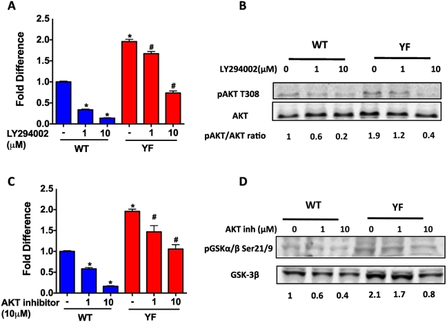
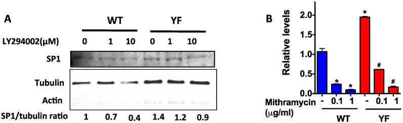
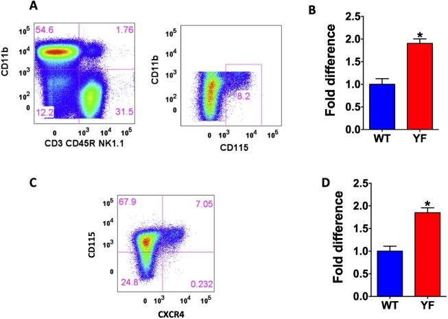
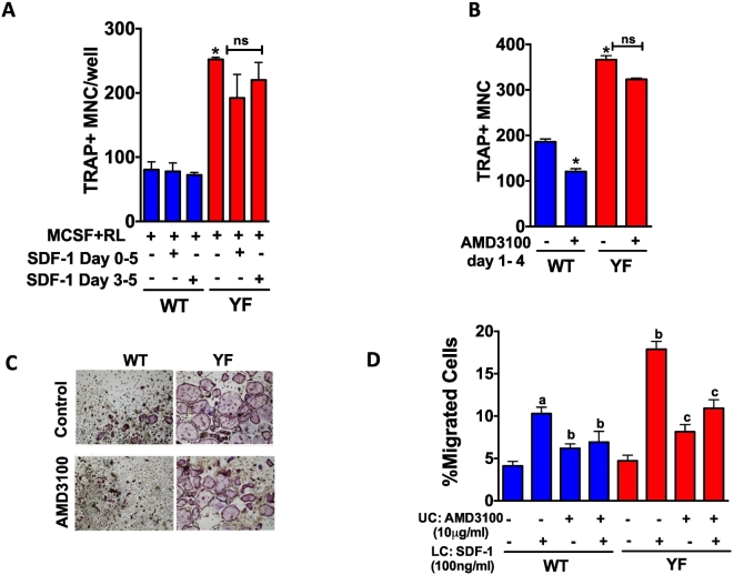
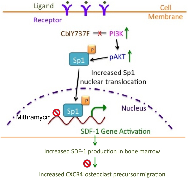
Our previous studies showed that in a mouse model in which PI3K-AKT activation was increased (YF mice), osteoclast numbers and levels of SDF-1, a chemokine, were augmented. The purpose of this study was to delineate the role of PI3K activation in regulating SDF-1 production and examine whether SDF-1 can stimulate differentiation and/or migration of osteoclast precursors. Using flow cytometric analysis, we demonstrated that compared to wild type mice, bone marrow of YF mice had increased numbers of CXCL12 abundant reticular (CAR) cells, that are a major cell type responsible for producing SDF-1. At the molecular level, transcription factor specificity protein 1 (Sp1) induced an increased transcription of SDF-1 that was dependent on PI3K/AKT activation. YF mice also contained an increased number of osteoclast precursors, in which expression of CXCR4, a major receptor for SDF-1, was increased. SDF-1 did not induce differentiation of osteoclast precursors into mature osteoclasts; compared to cells derived from WT mice, cells obtained from YF mice were more responsive to SDF-1. In conclusion, we demonstrate that PI3K activation resulted in increased SDF-1, increased the number of osteoclast precursors, and enhanced osteoclast precursor migration in response to SDF-1.

Keywords: CAR cells; Migration; Osteoclast precursors; PI3K; SDF-1.


Figures

Fig. 1
Fig. 1
Number of CXCL12 abundant reticular cells and SDF-1 production in YF bone marrow. Flow analysis of CAR cells and SDF-1 production. A. Total bone marrow cells were gated for a population negative for CD45, Ter119, CD31, and Sca1 and positive for VCAM and Pdgfr, the phenotype of CAR cells. B. Number of CAR cells in bone marrow of 8 week-old WT and YF male mice. Percentage of CAR cells in bone marrow was calculated as the number in YF bone marrow relative to WT bone marrow. Data is pooled from five independent experiments (WT, n = 25; YF n = 27). C. Intracellular level of SDF-1 was determined by the mean fluorescence intensity (factor of mean SDF-1 staining and SDF-1 positive cell count). Histogram in grey shade represents mouse IgG FITC isotype control. Bold black line represents SDF-1 FITC stain in YF cells and thin grey line represents in WT cells. D. SDF-1 intracellular level is shown as amount of protein in CAR cells in YF bone marrow relative to CAR cells in WT bone marrow (n = 9 mice/genotype). E. qRT-PCR analysis of SDF-1 mRNA levels in FACS sorted CAR cells from bone marrow (n = 9 mice/genotype). *P value of <0.05 vs. WT.
Fig. 2
Fig. 2
Increased PI3K activity and increased transcription of SDF-1 in the absence of Cbl-PI3K interaction. A. qRT-PCR analysis of SDF-1 mRNA levels in magnetically sorted CD45 Ter119 CD31 Sca1 negative population of bone marrow cells, cultured and treated with PI3K inhibitor LY294002 at indicated concentrations for 3 h. SDF-1 mRNA levels were normalized to GAPDH, and values shown are relative to the SDF-1 mRNA levels in untreated WT cells. B. Western blotting analysis of AKT phosphorylation, indicative of PI3K activity. Bands were quantified by LICOR odyssey software and the ratio of pAKT to total AKT at indicated concentrations of PI3K inhibitor, LY294002 is shown below the blots with values relative to untreated WT cells. C. qRT-PCR analysis of SDF-1 mRNA levels following treatment with an AKT inhibitor at the indicated concentrations D. Western blotting analysis of GSK phosphorylation, indicative of AKT activity. A representative of 3 independent experiments is shown and P value of <0.05 vs. WT; *compared to untreated WT cells, # compared to untreated YF cells.
Fig. 3
Fig. 3
Sp1 mediated transcription of SDF-1. A. Nuclear protein from cells treated with LY294002 for 3 h at different concentrations was subjected to Western blotting. Blot was probed with antibody for SP1. The blot was stripped and reprobed for actin (in cytoplasm) and tubulin (in nucleus) to show the relative absence of cytoplasmic proteins in the nuclear lysate. Sp1/tubulin ratio is shown below the Western blot. B. Cells were treated with Mithramycin (inhibits Sp1 binding to GC-rich DNA) at indicated concentrations for 3 h and SDF-1 mRNA levels were determined by qRT-PCR analysis. Values are shown as relative to untreated WT cells. * A representative of 3 independent experiments is shown and P value of <0.05 from student's t-test compared to untreated WT cells, # compared to untreated YF cells.
Fig. 4
Fig. 4
Absence of Cbl-PI3K interaction results in increased number of osteoclast precursors expressing SDF-1 receptor, CXCR4 in the bone marrow of mice. Flow analysis of osteoclast precursors in the bone marrow. A. Total bone marrow from 8 week old male mice was gated for CD3- CD45R- NK1.1- cells, which express low or no CD11b and high levels of CD115 (c-Fms) were considered osteoclast precursors. B. Percentage of cells was calculated as the number in YF tissues relative to WT, and data pooled from independent experiments is shown (WT, n = 25; YF, n = 28). C. CD3- CD45R- NK1.1- CD11b lo/- CD115 hi cells were further gated for the expression of SDF-1 receptor CXCR4. D. CXCR4 positive osteoclast precursor population is shown (WT, n = 13; YF, n = 14). Percentage of cells was calculated as the number in YF tissues relative to WT, and data pooled from independent experiments is shown. * P value of <0.05 vs. WT.
Fig. 5
Fig. 5
SDF-1 mediates increased migration but not differentiation of osteoclast precursors. Transmigration assay of osteoclast precursors in response to SDF-1. A. Osteoclast precursors were placed in the upper chamber of the transwell with or without AMD3100. SDF-1 was added to the lower chamber. Percentage of cells, which migrated to the lower chamber was quantified and data is expressed relative to WT cells without treatment in the absence of SDF-1 in the lower chamber. B. Osteoclast precursors were differentiated in the presence of MCSF (20 ng/ml) and RANKL (50 ng/ml) in the presence or absence of SDF-1 (100 ng/ml) added at indicated times. After 5 days of differentiation, cells were TRAP stained and TRAP positive cells with >3 nuclei were considered as osteoclasts. C. Osteoclast differentiation was performed in the presence or absence ofAMD3100 with SDF-1 treatment (100 ng/ml). Images of TRAP positive cells at 10× magnification are shown. D. Bar graph shows the number of multinucleated (>3 nuclei/cell) TRAP positive cells. In panel A, a: compared to untreated WT cells, b: compared to WT cells exposed to SDF-1 in lower chamber, c: compared to YF cells exposed to SDF-1 in lower chamber. In B, D * compared to WT cells without SDF-1 or AMD3100 treatment. A representative of 3 independent experiments is shown. * P value of <0.05 from ANOVA, Tukey's post hoc test was considered statistically significant.
Fig. 6
Fig. 6
Proposed model depicting the role of PI3K/AKT/Sp1 axis on SDF-1 expression in CAR cells and the effect of increased SDF-1 levels on osteoclast precursor migration in response to increased PI3K activation. Loss of Cbl-PI3K interaction results in increased PI3K activity, which leads to increased phosphorylation of AKT. Sp1, an important substrate of PI3K is activated, translocated to the nucleus and binds to the SDF-1 promoter regions to activate SDF-1 transcription in CAR cells. Sp1 binding to SDF-1 promoter regions is inhibited by Mithramycin treatment resulting in decreased SDF-1 transcription to a lesser extent in YF cells compared to wild type cells due to increased Sp1 activation in YF cells. Increased SDF-1 gene expression leads to increased SDF-1 protein levels, which stimulate migration of osteoclast precursors expressing SDF-1 receptor, CXCR4. AMD3100 blocks CXCR4 activation by SDF-1 and prevents osteoclast precursor migration, to a lesser extent in YF cells compared to wild type cells. Increased osteoclast precursor migration might lead to increased recruitment to the bone marrow milieu finally contributing to increased number of osteoclasts in YF mice lacking Cbl-PI3K interaction.

Similar articles

Cited by

References

    1. Adapala N.S., Barbe M.F., Langdon W.Y., Nakamura M.C., Tsygankov A.Y., Sanjay A. The loss of Cbl-phosphatidylinositol 3-kinase interaction perturbs RANKL-mediated signaling, inhibiting bone resorption and promoting osteoclast survival. J. Biol. Chem. 2010;285:36745–36758. - PMC - PubMed
    1. Adapala N.S., Barbe M.F., Langdon W.Y., Tsygankov A.Y., Sanjay A. Cbl-phosphatidylinositol 3 kinase interaction differentially regulates macrophage colony-stimulating factor-mediated osteoclast survival and cytoskeletal reorganization. Ann. N. Y. Acad. Sci. 2010;1192:376–384. - PubMed
    1. Adapala N.S., Barbe M.F., Tsygankov A.Y., Lorenzo J.A., Sanjay A. Loss of Cbl-PI3K interaction enhances osteoclast survival due to p21-Ras mediated PI3K activation independent of Cbl-b. J. Cell. Biochem. 2014;115:1277–1289. - PMC - PubMed
    1. Adapala N.S., Holland D., Scanlon V., Barbe M.F., Langdon W.Y., Tsygankov A.Y., Lorenzo J.A., Sanjay A. Loss of Cbl-PI3K interaction in mice prevents significant bone loss following ovariectomy. Bone. 2014;67:1–9. - PMC - PubMed
    1. Arai F., Miyamoto T., Ohneda O., Inada T., Sudo T., Brasel K., Miyata T., Anderson D.M., Suda T. Commitment and differentiation of osteoclast precursor cells by the sequential expression of c-Fms and receptor activator of nuclear factor kappaB (RANK) receptors. J. Exp. Med. 1999;190:1741–1754. - PMC - PubMed