. 2019 Apr 10;5(4):eaau7198.
doi: 10.1126/sciadv.aau7198. eCollection 2019 Apr.

VPS4 triggers constriction and cleavage of ESCRT-III helical filaments

Affiliations

VPS4 triggers constriction and cleavage of ESCRT-III helical filaments

Sourav Maity et al. Sci Adv. .

Abstract

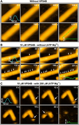
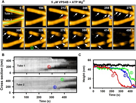
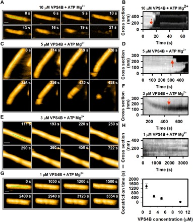
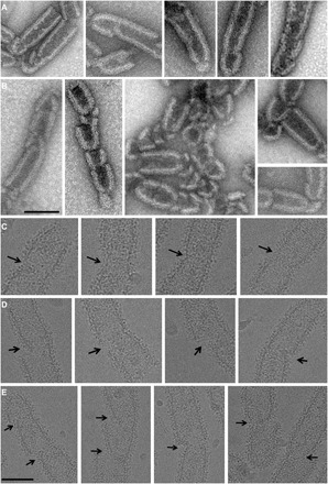
Many cellular processes such as endosomal vesicle budding, virus budding, and cytokinesis require extensive membrane remodeling by the endosomal sorting complex required for transport III (ESCRT-III). ESCRT-III protein family members form spirals with variable diameters in vitro and in vivo inside tubular membrane structures, which need to be constricted to proceed to membrane fission. Here, we show, using high-speed atomic force microscopy and electron microscopy, that the AAA-type adenosine triphosphatase VPS4 constricts and cleaves ESCRT-III CHMP2A-CHMP3 helical filaments in vitro. Constriction starts asymmetrically and progressively decreases the diameter of CHMP2A-CHMP3 tubular structure, thereby coiling up the CHMP2A-CHMP3 filaments into dome-like end caps. Our results demonstrate that VPS4 actively constricts ESCRT-III filaments and cleaves them before their complete disassembly. We propose that the formation of ESCRT-III dome-like end caps by VPS4 within a membrane neck structure constricts the membrane to set the stage for membrane fission.


Figures

Fig. 1
Fig. 1. Local deformation of VPS4B-treated CHMP2A-CHMP3 tubes and disassembly upon ATP hydrolysis.
(A) Clips of HS-AFM images of CHMP2A-CHMP3 tubes captured at 0.5 frame/s in the absence of VPS4B, showing no significant change in roughness of the tube surface. The inset represents the cross section (axial or radial) along the corresponding lines. (B) Clips of HS-AFM images captured at 1 frame/s, showing that the 10 μM VPS4-treated CHMP2A-CHMP3 tubes undergo discrete reductions of the tube radius (indicated with white arrows) without further structural changes in the absence of ATP Mg2+. The axial cross section at 231 s shows a clear reduction of at least 3 nm in the tube radius. Because of tip convolution, the exact value is likely higher than the observed result. (C) Clips of HS-AFM images captured at 1 frame/s from 10 μM VPS4-treated CHMP2A-CHMP3 tubes in presence of 200 μM ATP Mg2+. Upon ATP hydrolysis, CHMP2A-CHMP3 tubes disassembled within less than 1 min. White arrows show disassembly sites. Average tube height is 48 ± 3 nm (n = 30). Scale bars, 100 nm.
Fig. 2
Fig. 2. In presence of ATP Mg2+, VPS4B mediates local constriction (asymmetric) of the CHMP2A-CHMP3 tubes before disassembly.
(A) Clips of HS-AFM images captured at 0.5 frame/s, showing that the 5 μM VPS4B-treated CHMP2A-CHMP3 tubes undergo local constriction before disassembly. The constriction normally appears asymmetric (white arrows), i.e., the deformation starts from one side of the tube. Numbers in circles identify three constriction sites in the two tubes. Scale bar, 100 nm. (B) Kymographs of both tubes [green lines in (A)]. The progress of the constrictions (numbers in circles) is identifiable from the kymograph. (C) The height profiles of the tubes in (A) at nonconstricted site (in black curves) and at the constriction site over time derived from (A) and (B). Each color code is similar to the constriction sites (numbers in circles) in (A) and (B).
Fig. 3
Fig. 3. Effect of VPS4 concentration on CHMP2A-CHMP3 tube remodeling in presence of ATP Mg2+.
(A) Clips of HS-AFM images captured at 1 frame/s, showing a rapid disassembly of tube, treated with 10 μM VPS4B. (B) Kymograph taken from (A), showing the time and position of first constriction and/or disassembly (marked by red arrow). (C) Same as (A) but for 5 μM VPS4B-treated tubes. Frame rate, 1 frame/s. (D) Same as (B) but derived from (C). (E and F) Same as (C) and (D), respectively, but for 3 μM VPS4B-treated tubes. One can see that, at this concentration, the tube goes through constriction but not disassembly. (G and H) Same as (C) and (D), respectively, but for 1 μM VPS4B-treated tubes. Images in (G) show that at, 1 μM VPS4B concentration (with ATP Mg2+), the tube undergoes a partial constriction after a significant amount of time. Frame rate, 0.33 frame/s. (I) Effect of VPS4B concentration on the cleavage of CHMP2A-CHMP3 tubes. The cleavage time is considered from the moment of addition of 200 μM ATP Mg2+ until the first cleavage occurs (red arrows in the kymograph). For four different concentrations of VPS4B, more than 20 tubes were investigated. Scale bars, 100 nm.
Fig. 4
Fig. 4. EM imaging of CHMP2A-CHMP3 tube remodeling by VPS4B.
(A and B) Negative-stain images of CHMP2A-CHMP3 tubes incubated with 5 μM VPS4B and 200 μM ATP Mg2+, indicating (A) start sites of cleavage and (B) the generation of dome-like end caps. Scale bar, 100 nm. (C to E) Cryo-EM images of CHMP2A-CHMP3 tubes incubated with 5 μM VPS4B and 200 μM ATP Mg2+. (C) Images of early stages of constriction sites, (D) asymmetric constriction start sites, and (E) the generation of dome-like end caps. Scale bar, 100 nm.
Fig. 5
Fig. 5. Model of membrane constriction induced by remodeling of CHMP2A-CHMP3 filaments.
(A) CHMP2A-CHMP3 helical filaments assemble within a membrane neck structure such as a vesicle or virus bud or at the midbody. Currently, we do not know how many turns assemble in vivo. (B) VPS4 forms an asymmetric hexamer structure in the presence of ATP and Mg2+. This structure needs to assemble on functional ESCRT-III filaments, and ATP-driven rotation threads the substrate via its central pore. Because CHMP2A-CHMP3 polymer assembly is directional, the assembly of VPS4 acting clockwise or anticlockwise is likely to be important. The assembly of one VPS4 complex might be sufficient to induce CHMP2A-CHMP3 constriction, which often starts asymmetrically (middle). This can lead to membrane constriction and cleavage of the CHMP2A-CHMP3 filament. (C) Alternatively, two adjacent VPS4B complexes remodel CHMP2A-CHMP3 filaments acting clockwise and anticlockwise, thereby leading to the generation of two dome-like end caps and cleavage of the CHMP2A-CHMP3 filament. In both scenarios, constriction of CHMP2A-CHMP3 could prime the site for fission, and cleavage of the filament might play an important role in tension release as proposed (53).

References

    1. Henne W. M., Stenmark H., Emr S. D., Molecular mechanisms of the membrane sculpting ESCRT pathway. Cold Spring Harb. Perspect. Biol. 5, a016766 (2013). - PMC - PubMed
    1. McCullough J., Colf L. A., Sundquist W. I., Membrane fission reactions of the mammalian ESCRT pathway. Annu. Rev. Biochem. 82, 663–692 (2013). - PMC - PubMed
    1. Scourfield E. J., Martin-Serrano J., Growing functions of the ESCRT machinery in cell biology and viral replication. Biochem. Soc. Trans. 45, 613–634 (2017). - PubMed
    1. Juan T., Fürthauer M., Biogenesis and function of ESCRT-dependent extracellular vesicles. Semin. Cell Dev. Biol. 74, 66–77 (2018). - PubMed
    1. Stoten C. L., Carlton J. G., ESCRT-dependent control of membrane remodelling during cell division. Semin. Cell Dev. Biol. 74, 50–65 (2018). - PMC - PubMed

Publication types

MeSH terms

Substances

LinkOut - more resources