. 2019 Apr 10;5(4):eaav4340.
doi: 10.1126/sciadv.aav4340. eCollection 2019 Apr.

Targeting dePARylation selectively suppresses DNA repair-defective and PARP inhibitor-resistant malignancies

Affiliations

Targeting dePARylation selectively suppresses DNA repair-defective and PARP inhibitor-resistant malignancies

Shih-Hsun Chen et al. Sci Adv. .

Abstract

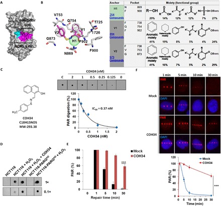
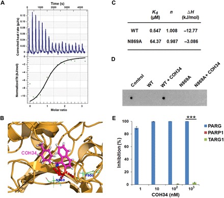
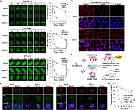
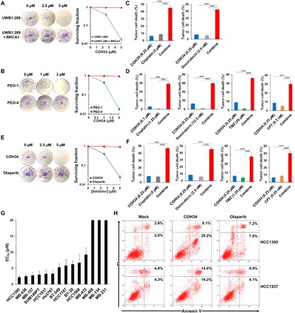
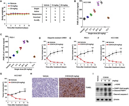
While poly(ADP-ribosyl)ation (PARylation) plays an important role in DNA repair, the role of dePARylation in DNA repair remains elusive. Here, we report that a novel small molecule identified from the NCI database, COH34, specifically inhibits poly(ADP-ribose) glycohydrolase (PARG), the major dePARylation enzyme, with nanomolar potency in vitro and in vivo. COH34 binds to the catalytic domain of PARG, thereby prolonging PARylation at DNA lesions and trapping DNA repair factors. This compound induces lethality in cancer cells with DNA repair defects and exhibits antitumor activity in xenograft mouse cancer models. Moreover, COH34 can sensitize tumor cells with DNA repair defects to other DNA-damaging agents, such as topoisomerase I inhibitors and DNA-alkylating agents, which are widely used in cancer chemotherapy. Notably, COH34 also efficiently kills PARP inhibitor-resistant cancer cells. Together, our study reveals the molecular mechanism of PARG in DNA repair and provides an effective strategy for future cancer therapies.


Figures

Fig. 1
Fig. 1. COH34 is a potent and cell-active PARG inhibitor.
(A) Predicted inhibitor site (magenta area) of PARG. (B) Site-moiety map and docked conformation of COH34 (magenta) in inhibitor site. (C) Chemical structure and formula of COH34. The IC50 value of COH34 was measured by dot blotting with PAR antibody in a dose course of COH34 (n = 3 independent experiments). (D and E) HCT116 cells were pretreated with or without COH34 (0.1 μM) for 1 hour before treatment with 0.5 mM H2O2 at 37°C for 15 min. HCT116 cells without H2O2 treatment and HCT116-PARG knockdown (HCT116-PARGKD) cells with H2O2 treatment are negative control and positive control, respectively. The extent of PAR was determined by dot blotting with anti-PAR antibody. The time course data are shown in the histograms from three independent experiments. ***P < 0.001. (F) A microscope-coupled laser scissors system was used to generate DNA damage in nucleus. PAR at DNA lesions in U2OS cells with or without 100 nM PARG inhibitor (COH34) treatment was immunostained with PAR antibody (red dots) after laser scissors. The kinetics of the accumulation of PAR at DNA damage sites in a time course was shown as mean ± SD from 50 cells (n = 3 independent experiments). ***P < 0.001.
Fig. 2
Fig. 2. COH34 specifically binds to the catalytic site of PARG.
(A) The affinity between COH34 and the recombinant catalytic domain of PARG was measured by ITC. Titration of COH34 into a solution containing the purified protein was performed at 25°C using a Nano ITC instrument. The binding isotherm shows the fit of the data to an equilibrium-binding isotherm. The fit provides an equilibrium Kd for the binding of COH34 to the catalytic domain of PARG. (B) A predicted binding mode of COH34 at the catalytic site of PARG (PDB: 4BLI). COH34 (magenta) fits well in the catalytic domain (orange) and interacts with two residues (green), N869 and F900, through hydrogen bonds (red dots). (C) ITC parameters between the wild-type PARG (WT) or the N869A mutant and COH34 are summarized in the table. (D) The N869A mutant retains the enzymatic activity, and COH34 is unable to suppress the N869A mutant. PAR digestion assays were performed with or without COH34 (10 nM). Results were analyzed using dot blotting with anti-PAR antibody (n = 3 independent experiments). Control means PAR only. (E) Target selectivity assay was carried out using PARG, PARP1, and TARG1 with indicated concentrations of COH34. COH34 against PARG and PARP1 activity was analyzed by dot blotting with anti-PAR antibody. TARG1 inhibition results were determined by Western blot with anti–ADP-ribose antibody. Average inhibition of targets in a dose course of COH34 is shown in the histograms (n = 3 independent experiments). ***P < 0.001
Fig. 3
Fig. 3. COH34-dependent trapping mechanism affects DNA damage repair.
(A) The relocation kinetics of XRCC1, CHFR, and APLF to DNA damage sites. GFP-XRCC1, CHFR, or APLF was expressed in U2OS cells. Cells were with or without pretreatment of 0.1 μM COH34 for 1 hour, and the relocation kinetics was monitored in a time course following laser microirradiation. Results are shown as mean ± SD from 50 cells (n = 3 independent experiments). ***P < 0.001. (B and C) KillerRed is a light-induced system that generates reactive oxygen species–driven DNA damage in cells. Cells expressing KillerRed protein were pretreated with or without COH34 (0.1 μM) for 1 hour before treatment with white light at 25°C for 10 min. KillerRed signal (DNA damage site, red dot) was detected with KillerRed antibody following light treatment. The relocation kinetics of endogenous PAR and XRCC1 was examined with PAR and XRCC1 antibodies. (D) The relocation kinetics results are summarized and presented as mean ± SD from 50 cells (n = 3 independent experiments). ***P < 0.001. (E) Time course of the accumulation of γ-H2AX foci after IR (1 Gy) treatment. U2OS cells were with or without pretreatment of COH34 (0.1 μM) for 1 hour, and the γ-H2AX foci were analyzed in a time course following IR treatment. Immunofluorescence was performed with γ-H2AX antibody. (F) Scheme of suppression of PARG traps DNA damage repair factors at the vicinity of DNA lesions and affects DNA damage repair.
Fig. 4
Fig. 4. COH34 selectively kills BRCA-mutant and PARP inhibitor–resistant cancer cells.
(A and B) Colony formation assays of the BRCA1-mutant ovarian cancer cells (UWB1.289) and BRCA2-mutant ovarian cancer cells (PEO-1) following 14 days of treatment with DMSO and indicated concentrations of COH34. BRCA1/2-reconstituted cells (UWB1.289 + BRCA1 and PEO-4) are used as controls. Cells were stained with crystal violet. Average cell viability is presented as mean ± SD. (C and D) Combination treatments with a series of concentrations of PARG inhibitor COH34 and DNA-damaging agents (cisplatin, doxorubicin, temozolomide, or camptothecin) in UWB1.289 or PEO-1 cells. The combination condition shows the best synergistic effect to kill cancer cells, and data are shown in the histograms. ***P < 0.001. (E) Colony formation assay of the olaparib-resistant BRCA1-mutant ovarian cancer cells (SYr12) following 14 days of treatment with DMSO and indicated concentrations of COH34 or olaparib. Cells were stained with crystal violet. Average cell viability is shown as mean ± SD. (F) Combination treatments with a series of concentrations of PARG inhibitor (COH34) and DNA-damaging agents [cisplatin, doxorubicin, temozolomide (TMZ), or camptothecin (CPT)] in SYr12 cells. The combination condition, which is the best synergistic effect to kill cancer cells, is shown in the histograms. ***P < 0.001. (G) Cell viability assay in a panel of triple-negative breast cancer (TNBC) cell lines treated with 0.625 to 20 μM COH34 at 37°C for 14 days. The average EC50 value of each cell line is presented as mean ± SD. (H) Annexin V/PI apoptosis analyses of HCC1395 and HCC1937 cells following COH34 or olaparib treatment. Cells were treated with 5 μM PARG inhibitor COH34 or 10 μM olaparib at 37°C for 72 hours and then collected for annexin V and PI staining. Results were analyzed by flow cytometry. The percentages of annexin V–positive (lower right quadrant) and annexin V/PI–double-positive (upper right quadrant) cells from these experiments are shown in the relevant quadrants. (A to H) Data were from three independent experiments.
Fig. 5
Fig. 5. COH34 exhibits antitumor activity in PARP inhibitor–resistant and DNA damage repair–defective cell line xenografts.
(A) Toxicity assay of COH34 in mice: Female NSG adult mice were dosed intraperitoneally with 30% solutol (vehicle, n = 3) or COH34 in 30% solutol at 10 mg/kg (n = 3) or 20 mg/kg (n = 3) once daily for 10 days. They were weighed daily and observed for signs of pain and distress. (B and C) PAR analyses of tumor samples taken from NSG mice of HCC1395 and PEO-1 xenografts by dot blotting. Samples were taken after a single-dose treatment at the indicated time from each of the five mice receiving either COH34 (20 mg/kg) or vehicle dosed intraperitoneally. (D) Effect of COH34 in an olaparib-resistant UWB1.289 xenograft. Eight-week-old female NSG mice were injected with 8 million SYr12 cells. After tumors reached an average size of ~70 mm3, they were treated with vehicle (n = 6) or COH34 (20 mg/kg, n = 6) through intraperitoneal injections for 2 weeks. ***P < 0.001. (E) Effect of COH34 in a BRCA2-mutant ovarian cancer xenograft. Eight-week-old female NSG mice were injected with 10 million PEO-1 cells. Mice were randomized into two treatment groups of six mice. Mice were treated with COH34 (20 mg/kg) or vehicle intraperitoneally once daily for 2 weeks when tumors reached an average size of ~90 mm3. ***P < 0.001. (F and G) Effect of COH34 in BRCA-mutant TNBC xenografts. Eight-week-old female NSG mice were injected with 8 million HCC1395 or HCC1937 cells. After tumors reached an average size of ~85 mm3, they were treated with vehicle (14 days of treatment, n = 6) or COH34 (20 mg/kg, 14 days of treatment; n = 6) through intraperitoneal injections. ***P < 0.001. (H) Analysis of apoptosis, detected by immunohistochemistry. Shown are images from representative sections of tumor samples from COH34 and vehicle-treated mice. (I) Lysates of HCC1395 xenografts (~60 mg) from COH34 and vehicle-treated mice were subjected to Western blotting with γ-H2AX, PARP1, cleaved caspase-3, or β-Actin antibody. β-Actin was used as a control of protein loading.

Similar articles

Cited by

References

    1. Kraus W. L., PARPs and ADP-ribosylation: 50 years ... and counting. Mol. Cell 58, 902–910 (2015). - PMC - PubMed
    1. Bürkle A., Poly(ADP-ribose)—The most elaborate metabolite of NAD. FEBS J. 272, 4576–4589 (2005). - PubMed
    1. Hassa P. O., Haenni S. S., Elser M., Hottiger M. O., Nuclear ADP-ribosylation reactions in mammalian cells: where are we today and where are we going? Microbiol. Mol. Biol. Rev. 70, 789–829 (2006). - PMC - PubMed
    1. Vyas S., Matic I., Uchima L., Rood J., Zaja R., Hay R. T., Ahel I., Chang P., Family-wide analysis of poly(ADP-ribose) polymerase activity. Nat. Commun. 5, 4426 (2014). - PMC - PubMed
    1. Leung A. K. L., Poly(ADP-ribose): An organizer of cellular architecture. J. Cell Biol. 205, 613–619 (2014). - PMC - PubMed

Publication types

Substances

LinkOut - more resources