Review
. 2019 Jun;286(12):2240-2260.
doi: 10.1111/febs.14854. Epub 2019 Apr 29.

Posttranslational modifications of titin from cardiac muscle: how, where, and what for?

Affiliations
Review

Posttranslational modifications of titin from cardiac muscle: how, where, and what for?

Franziska Koser et al. FEBS J. 2019 Jun.

Abstract

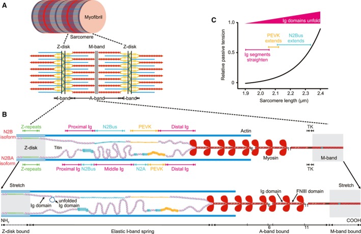
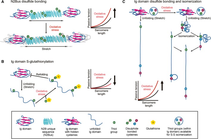
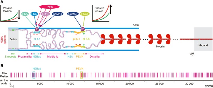
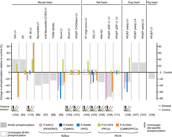
Titin is a giant elastic protein expressed in the contractile units of striated muscle cells, including the sarcomeres of cardiomyocytes. The last decade has seen enormous progress in our understanding of how titin molecular elasticity is modulated in a dynamic manner to help cardiac sarcomeres adjust to the varying hemodynamic demands on the heart. Crucial events mediating the rapid modulation of cardiac titin stiffness are post-translational modifications (PTMs) of titin. In this review, we first recollect what is known from earlier and recent work on the molecular mechanisms of titin extensibility and force generation. The main goal then is to provide a comprehensive overview of current insight into the relationship between titin PTMs and cardiomyocyte stiffness, notably the effect of oxidation and phosphorylation of titin spring segments on titin stiffness. A synopsis is given of which type of oxidative titin modification can cause which effect on titin stiffness. A large part of the review then covers the mechanically relevant phosphorylation sites in titin, their location along the elastic segment, and the protein kinases and phosphatases known to target these sites. We also include a detailed coverage of the complex changes in phosphorylation at specific titin residues, which have been reported in both animal models of heart disease and in human heart failure, and their correlation with titin-based stiffness alterations. Knowledge of the relationship between titin PTMs and titin elasticity can be exploited in the search for therapeutic approaches aimed at softening the pathologically stiffened myocardium in heart failure patients.

Keywords: cytoskeleton; elasticity; heart; heart disease; oxidation; oxidative stress; phosphorylation; protein kinase; protein phosphatase.


Conflict of interest statement

The authors declare no conflict of interest.

Figures

Figure 1
Figure 1
Titin isoforms and force‐extension mechanisms in human cardiac sarcomeres. (A) Schematic of a section of a myofibril constituted by sarcomeres (bordered by Z‐disks), which consist mainly of the three myofilaments, actin (thin), myosin (thick), and titin (elastic). (B) A half‐sarcomere is shown at two different stretch states. Cardiac titin isoforms, N2B and N2BA, are drawn as being coexpressed in the half‐sarcomere; molecular spring elements within elastic I‐band titin are highlighted. TK, titin kinase domain. (C) Relative titin‐based passive tension vs. sarcomere length relationship of a cardiomyocyte. Colors indicate the sequential extension of I‐band titin segments of the N2B isoform, which includes initial straightening of the Ig domain regions, followed by extension of the PEVK and N2Bus elements, and the continuous increase in the probability of Ig domain unfolding with stretching.
Figure 2
Figure 2
Mechanisms of titin‐based passive tension modulation by oxidative stress‐induced titin modifications. (A) Formation of up to three intramolecular disulphide bonds within the human N2Bus element of titin under oxidative stress increases titin‐based passive tension in cardiomyocytes. (B) Ig domain unfolding due to sarcomere stretching causes exposure of hidden (‘cryptic’) cysteines in Ig domains, which can become S‐glutathionylated under oxidative conditions. S‐glutathionylation prevents Ig domain refolding, resulting in decreased titin‐based passive tension. (C) Isomerization of disulfide bonds of the cysteine triad in titin Ig domains can occur under oxidative conditions. Depending on where the intramolecular S‐S cross‐linking occurs, titin‐based passive tension increases by different amplitudes. The key on bottom explains the different shapes and colors shown in the figure.
Figure 3
Figure 3
Potential and verified phosphorylation sites in human titin. (A) Layout of the N2BA titin isoform in a cardiac half‐sarcomere, highlighting protein kinases (for details, see main text) and protein phosphatase (PP)5 known to mediate phosphorylation/ dephosphorylation at two distinct molecular spring elements, N2Bus and constitutively expressed PEVK (light green bit of PEVK). Phosphorylation of N2Bus reduces titin‐based passive tension, whereas phosphorylation of PEVK increases it, which is explained by the different net charge of these elements. Constitutive PEVK has a net positive charge (+) and high isoelectric point (pI), N2Bus a net negative charge (−) and low pI. Note that the alternatively spliced PEVK subsegment (yellow bit of PEVK) also has a net negative charge. (B) Known potential phosphosites in human cardiac titin (vertical red bars), from http://www.phosphosite.org 85. Locations of phosphosites verified by site‐specific methods are highlighted (blue and green boxes). TK, titin kinase domain.
Figure 4
Figure 4
Changes in titin phosphorylation in animal models of heart disease and relationship with alterations in cardiomyocyte passive tension. The size of the bars indicates the relative amount of change in phosphorylation in diseased hearts vs. the respective control, healthy hearts (for some conditions, an average value is shown). The relative changes are as indicated by the respective study authors and therefore, magnitude comparisons between studies may not be plausible. Note the heterogeneity in the direction of change in all‐titin phosphorylation among the different models. Also, note that in failing hearts, the N2Bus element is frequently hypophosphorylated at one or more sites, whereas phosphoserine (S11878) within the PEVK element is usually hyperphosphorylated. Conversely, PEVK site S12022 mostly shows hypophosphorylation. For those studies where titin‐based passive tension was measured, the direction of change (arrow) in disease (red curves) relative to healthy heart samples (green curves) is indicated. Protein kinases known to target individual phosphosites are listed in parentheses. References are in square brackets. TAC, transversal aortic constriction (afterload increase); I/R, ischemia/reperfusion injury; MI, myocardial infarction; T2DM, type 2 diabetes mellitus; Shunt, aorto‐caval shunt model (preload increase); HFpEF, heart failure with preserved ejection fraction; TtnD/Ajxn, deletion of titin segment at I‐band/A‐band junction; HT, hypertension; PAH, pulmonary arterial hypertension; ZSF‐1, Zucker spontaneously hypertensive, fatty‐1 model; LV, left ventricle; RV, right ventricle; LA, left atrium.
Figure 5
Figure 5
Changes in titin phosphorylation in human failing hearts and relationship with alterations in cardiomyocyte passive tension. The size of the bars indicates the relative amount of change in phosphorylation in failing vs. nonfailing hearts (for some conditions, an average value is shown). The relative changes are as indicated by the respective study authors and therefore, magnitude comparisons between studies may not be plausible. Note the absence of hyperphosphorylation of all‐titin. Also, note the general pattern of hypophosphorylation at one or more N2Bus sites and hyperphosphorylation at PEVK site S11878, whereas phosphorylation at S12022 mostly remains unaltered. For those studies where titin‐based passive tension was measured, the direction of change (arrow) in disease (red lines) relative to healthy heart samples (green lines) is indicated. Protein kinases known to target individual phosphosites are listed in parentheses. References are in square brackets. DCM, dilated cardiomyopathy; CHF, congestive heart failure; IDCM, idiopathic DCM; HCM, hypertrophic cardiomyopathy; HFpEF, heart failure with preserved ejection fraction; HFrEF, heart failure with reduced ejection fraction; HT, hypertensive heart disease; PAH, pulmonary arterial hypertension; AS, aortic stenosis; PPCM, peripartum cardiomyopathy; ISHD, ischemic heart disease. LV, left ventricle; RV, right ventricle.

References

    1. Anderson BR & Granzier HL (2012) Titin‐based tension in the cardiac sarcomere: molecular origin and physiological adaptations. Prog Biophys Mol Biol 110, 204–217. - PMC - PubMed
    1. Tskhovrebova L & Trinick J (2010) Roles of titin in the structure and elasticity of the sarcomere. J Biomed Biotechnol 2010, 612482. - PMC - PubMed
    1. Linke WA (2008) Sense and stretchability: the role of titin and titin‐associated proteins in myocardial stress‐sensing and mechanical dysfunction. Cardiovasc Res 77, 637–648. - PubMed
    1. Robison P, Caporizzo MA, Ahmadzadeh H, Bogush AI, Chen CY, Margulies KB, Shenoy VB & Prosser BL (2016) Detyrosinated microtubules buckle and bear load in contracting cardiomyocytes. Science 352, aaf0659. - PMC - PubMed
    1. Chen CY, Caporizzo MA, Bedi K, Vite A, Bogush AI, Robison P, Heffler JG, Salomon AK, Kelly NA, Babu A, et al (2018) Suppression of detyrosinated microtubules improves cardiomyocyte function in human heart failure. Nat Med 24, 1225–1233. - PMC - PubMed

Publication types