. 2019 Apr 16;5(10):e125665.
doi: 10.1172/jci.insight.125665.

Cullin-3 dependent deregulation of ACTN1 represents a new pathogenic mechanism in nemaline myopathy

Affiliations

Cullin-3 dependent deregulation of ACTN1 represents a new pathogenic mechanism in nemaline myopathy

Jordan Blondelle et al. JCI Insight. .

Abstract

Nemaline myopathy is a congenital neuromuscular disorder characterized by muscle weakness, fiber atrophy and presence of nemaline bodies within myofibers. However, the understanding of underlying pathomechanisms is lacking. Recently, mutations in KBTBD13, KLHL40 and KLHL41, three substrate adaptors for the E3-ubiquitin ligase Cullin-3, have been associated with early-onset nemaline myopathies. We hypothesized that deregulation of Cullin-3 and its muscle protein substrates may be responsible for the disease development. Using Cullin-3 knockout mice, we identified accumulation of non-muscle alpha-Actinins (ACTN1 and ACTN4) in muscles of these mice, which we also observed in KBTBD13 patients. Our data reveal that proper regulation of Cullin-3 activity and ACTN1 levels is essential for normal muscle and neuromuscular junction development. While ACTN1 is naturally downregulated during myogenesis, its overexpression in C2C12 myoblasts triggered defects in fusion, myogenesis and acetylcholine receptor clustering; features that we characterized in Cullin-3 deficient mice. Taken together, our data highlight the importance for Cullin-3 mediated degradation of ACTN1 for muscle development, and indicate a new pathomechanism for the etiology of myopathies seen in Cullin-3 knockout mice and nemaline myopathy patients.

Keywords: Mouse models; Muscle Biology; Skeletal muscle; Ubiquitin-proteosome system.


Conflict of interest statement

Conflict of interest: The authors have declared that no conflict of interest exists.

Figures

Figure 1
Figure 1. Deletion of Cullin-3 in skeletal muscles affects the ubiquitin-proteasome system.
(A) Immunoblot analysis showing loss of Cullin-3 (Cul3) protein only in skeletal muscle (Sk. muscle) of E18.5 skm-KO mice. (B) Quantification of Cullin-3 protein levels in E18.5 skeletal muscles (n = 3 for each genotype). *P < 0.05 by 2-tailed t test. ctl, control. (C) Immunoblot analysis showing a decrease in NEDD8-associated proteins in E18.5 skeletal muscles of skm-KO mice. (D) Quantification of 80-kDa NEDD8-associated protein levels in E18.5 skeletal muscles (n = 3 for each genotype). **P < 0.01 by 2-tailed t test. (E) Immunoblot analysis showing a decrease in low molecular weights of K48-ubiquitin–associated proteins [Ubiq. (K48)] and no change in p62 expression levels in skeletal muscles of E18.5 skm-KO mice.
Figure 2
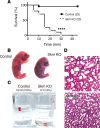
Figure 2. Loss of Cullin-3 during skeletal muscle development leads to postnatal death and respiratory defects.
(A) Survival curve of E18.5 embryos following C-section (n = 23 for control [ctl] and n = 20 for skm-KO). ****P < 0.0001 by log-rank (Mantel-Cox) and Gehan-Breslow-Wilcoxon tests. (B) Representative pictures of E18.5 embryos showing cyanosis and kyphosis of skm-KO mice. (C) Floating assay using lungs extracted from E18.5 skm-KO embryos and controls 5 minutes after the C-section and placed at the surface of water (n = 18 for ctl and n = 13 for skm-KO). (D) Cross section of E18.5 lungs 5 minutes after C-section stained with H&E, revealing collapsed alveoli in skm-KO embryos.
Figure 3
Figure 3. Absence of Cullin-3 leads to severe skeletal muscle myopathy.
(A) Body weight analysis of E18.5 embryos (n = 43 for ctl, n = 79 for heterozygous [f/+;cre+], and n = 41 for skm-KO). ****P < 0.001 by ANOVA and Bonferroni’s multiple-comparisons test. (B) Diaphragm weight analysis, revealing strong muscle atrophy of E18.5 skm-KO embryos. n = 10 for ctl, n = 19 for heterozygous (f/+;cre+), and n = 8 for skm-KO. ***P < 0.0001 by ANOVA and Bonferroni’s multiple-comparisons test. (C) Cross section of E18.5 diaphragms stained with H&E showing thinner muscle in skm-KO. Scale bar: 1 mm. (D) Immunoblot analysis showing a decrease in expression of muscle maturation markers in skm-KO diaphragms (n = 3 for each genotype). (E) Immunofluorescence staining of diaphragm myofibers with muscle ACTN2 and ACTN3 antibodies as well as DAPI. Arrowheads indicate centralized nuclei. Scale bar: 20 μm.
Figure 4
Figure 4. Loss of Cullin-3 leads to muscle fiber hypotrophy in vivo and myoblast fusion defect in vitro.
(A) Immunofluorescence staining of E18.5 diaphragms with WGA and DAPI revealing hypotrophy of the myofibers of skm-KO embryos. Arrowheads indicate centralized nuclei. Scale bar: 100 μm. (B) Distribution of fibers constituting diaphragms of E18.5 ctl and skm-KO depending on their cross-sectional area (CSA in μm2) (n = 3 embryos for each genotype and >11,554 fibers per genotype). (C) RT-PCR analysis of Cullin-3 and Cyclophilin B (CycloB) in satellite cells isolated from E18.5 ctl and skm-KO diaphragms over 3 days of differentiation in culture. Pro, proliferation; D1–D3, differentiation days 1–3. (D) Immunofluorescence of satellite cells fixed after 3 days of differentiation and stained with MyHC and β-actin antibodies. Scale bar: 100 μm. (E) Immunoblots of Cullin-3, MyHC, and β-actin on C2C12 myotubes transfected with an siRNA against Cullin-3 or a scrambled siRNA, showing efficient knockdown. (F) Fusion index (number of nuclei per myotube) of C2C12 cells transfected with a Cullin-3 or a scrambled siRNA and differentiated for 5 days (n = 3 per condition and >144 myotubes analyzed per experiment). *P < 0.05 by 2-tailed t test.
Figure 5
Figure 5. Diaphragm muscle is a hot spot for Cullin-3 substrate adaptors, and depletion of Cullin-3 leads to deregulation of protein levels.
(A) Immunoblot analysis showing the pattern of expression of Cullin-1, Cullin-3, and select Cullin-3 substrate adaptors in various mouse skeletal muscles. Myomesin-3 (Myom3) (86) was used as fiber-type marker. Gas, gastrocnemius; Sth, sternohyoideus; Pem, pectoralis major; Dia, diaphragm; Io, internal oblique; Pei, pectoralis minor; Mas, massester; Tri, triceps, Bib, biceps brachii; Cla, clavotrapezius; Edl, extensor digitorum longus; TA, tibialis anterior; Ste, sternomastoideus; Sol, soleus. (B) Volcano plot of all identified deregulated proteins in E18.5 diaphragms of skm-KO mice following iTRAQ mass spectrometry (3 diaphragms were pooled per sample, and 3 samples for each genotype were analyzed). (C) Volcano plot highlighting deregulated proteins involved in actin cytoskeleton modulation as well as proteins related to muscle and calcium handling (3 diaphragms were pooled per sample, and 3 samples for each genotype were analyzed).
Figure 6
Figure 6. Loss of Cullin-3 induces accumulation of ACTN1.
(A) Immunoblot analysis showing accumulation of ACTN1 in diaphragms of skm-KO embryos. (B) Quantification of ACTN1 protein levels in E18.5 diaphragms of ctl and skm-KO mice (n = 3 embryos for each genotype). *P < 0.05 by 2-tailed t test. (C) Immunoblots showing accumulation of ACTN1 and deregulation of sarcomeric myosin in satellite cells of skm-KO embryos after 3 days of differentiation. (D) Quantification of ACTN1 protein and myosin levels in satellite cells of ctl and skm-KO embryos after 3 days of differentiation. n = 3 technical replicates for each genotype. *P < 0.05, **P < 0.01 by 2-tailed t test. (E) Immunoblot analysis showing the specific loss of Cullin-3 in skeletal muscles of iCul3-KO (doxycycline-inducible Cullin-3–knockout) mice. Dotted line indicates that samples for hearts and soleus have been run on the same gel but not in adjacent lanes. (F) Immunoblot analysis showing accumulation of ACTN1, but not sarcomeric ACTN2, in skeletal muscles of iCul3-KO mice treated with doxycycline.
Figure 7
Figure 7. Overexpression of ACTN1 in muscle cells leads to differentiation defects.
(A) Immunoblot analysis showing overexpression of ACTN1 in C2C12 cells overexpressing the HA-ACTN1 construct. Pro, proliferation; D5, differentiation day 5. (B) Immunofluorescence staining of HA- (ctl) and HA-ACTN1–overexpressing myotubes with sarcomeric myosin antibody and DAPI after 5 days of differentiation. Scale bar: 200 μm. (C) Fusion index of cells expressing HA (ctl) and HA-ACTN1 constructs 5 days after differentiation, showing a decrease in the number of nuclei per myotube (n = 3 for each condition). **P < 0.01 by 2-tailed t test. (D) Distribution of myotubes depending on the number of nuclei (n = 3 for each condition).
Figure 8
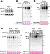
Figure 8. Cullin-3 protein levels and Cullin-RING ligase activity are increased over AchR clustering in vitro.
(A) Immunoblot analysis showing increased expression of Cullin-3 protein levels in C2C12 cells stimulated with neural agrin (0.5 μg/ml) for 48 hours in order to trigger acetylcholine receptor (AchR) clustering. (B) Immunoblot analysis showing increased neddylated proteins in C2C12 cells stimulated with agrin for 48 hours. (C) Immunoblot analysis showing increased levels of polyubiquitylated proteins (K48-linked) in C2C12 myotubes stimulated with agrin for 48 hours. (D) Immunoblot analyses showing a decrease in both Nedd8-linked and polyubiquitylated proteins in C2C12 myotubes stimulated with agrin and treated with MLN4924 compared with DMSO.
Figure 9
Figure 9. Cullin-3 is required for normal neuromuscular junction formation and acetylcholine receptor clustering.
(A and B) Immunofluorescence staining of the pre- and postsynaptic elements of E18.5 neuromuscular junctions (NMJs) in ctl and skm-KO diaphragms with antibodies against neurofilament and synaptophysin (NF/Syn) and fluorescent bungarotoxin (BGTX), showing (A) increased area of the motor endplate, dispersion of acetylcholine receptor (AchR) clusters across the diaphragm, and hyperarborization of the motoneuron in skm-KO embryos; but (B) normal innervation of the NMJ. Scale bars: 100 (A) μm; 20 μm (B). (C) Quantification of NMJ endplate widths (left panel; box-and-whiskers plot showing minimum to maximum values; + indicates average) and innervated AchRs (right panel) in the diaphragms of ctl and skm-KO embryos (n = 3 embryos for each genotype, >895 AchRs per genotype). ****P < 0.0001, by 2-tailed t test (left panel); P = 0.211 by 2-tailed t test (right panel). (D) Distribution of AchR clusters of ctl and skm-KO diaphragms according to their area (n = 3 embryos for each genotype). (E) Distribution of AchR cluster areas in 5-day-differentiated myotubes expressing HA (ctl) or HA-ACTN1 (n = 3 per condition and >817 AchR clusters were analyzed per condition).
Figure 10
Figure 10. Specific accumulation of non-muscle α-actinins in muscle tissues of patients with Cullin-3–related nemaline myopathies.
(A) Gomori trichrome staining of skeletal muscle cross sections from a healthy individual and a patient with a mutation in KBTBD13. (B) Immunoblot analyses of α-actinin isoforms expression levels in skeletal muscle biopsies from nemaline patients (NM patients; lane 1: TPM2 mutation; lane 2: NEB mutation), patients with mutations in KBTBD13 (lanes 3 and 4), and healthy individuals (lanes 5 and 6). Specific accumulation of non-muscle α-actinins (ACTN1 and ACTN4) was observed in samples from patients with mutations in KBTBD13. Muscle α-actinins (ACTN2 and ACTN3) did not show abnormal accumulation. Porin and Ponceau stains are shown as loading controls. (C) Immunofluorescence staining of cross sections from muscle biopsies of a healthy individual and a patient with a mutation in KBTBD13, revealing positive staining of nemaline bodies with non-muscle α-actinins (ACTN1 and ACTN4). Scale bars: 100 μm (A and C).
Figure 11
Figure 11. Model for the role of Cullin-3 during muscle fiber development.
S. Adaptor, substrate adaptor; Rbx1, RING-box protein 1; AchR, acetylcholine receptor; Ub, Ubiquitin; N8, Nedd8.

References

    1. Muscaritoli M, Lucia S, Molfino A, Cederholm T, Rossi Fanelli F. Muscle atrophy in aging and chronic diseases: is it sarcopenia or cachexia? Intern Emerg Med. 2013;8(7):553–560. doi: 10.1007/s11739-012-0807-8. - DOI - PubMed
    1. North K. What’s new in congenital myopathies? Neuromuscul Disord. 2008;18(6):433–442. doi: 10.1016/j.nmd.2008.04.002. - DOI - PubMed
    1. Bonaldo P, Sandri M. Cellular and molecular mechanisms of muscle atrophy. Dis Model Mech. 2013;6(1):25–39. doi: 10.1242/dmm.010389. - DOI - PMC - PubMed
    1. Petroski MD, Deshaies RJ. Function and regulation of cullin-RING ubiquitin ligases. Nat Rev Mol Cell Biol. 2005;6(1):9–20. - PubMed
    1. Bosu DR, Kipreos ET. Cullin-RING ubiquitin ligases: global regulation and activation cycles. Cell Div. 2008;3:7. - PMC - PubMed

Publication types

MeSH terms