. 2019 Apr 16;129(7):2872-2877.
doi: 10.1172/JCI124804.

Ectonucleotidase tri(di)phosphohydrolase-1 (ENTPD-1) disrupts inflammasome/interleukin 1β-driven venous thrombosis

Affiliations

Ectonucleotidase tri(di)phosphohydrolase-1 (ENTPD-1) disrupts inflammasome/interleukin 1β-driven venous thrombosis

Vinita Yadav et al. J Clin Invest. .

Abstract

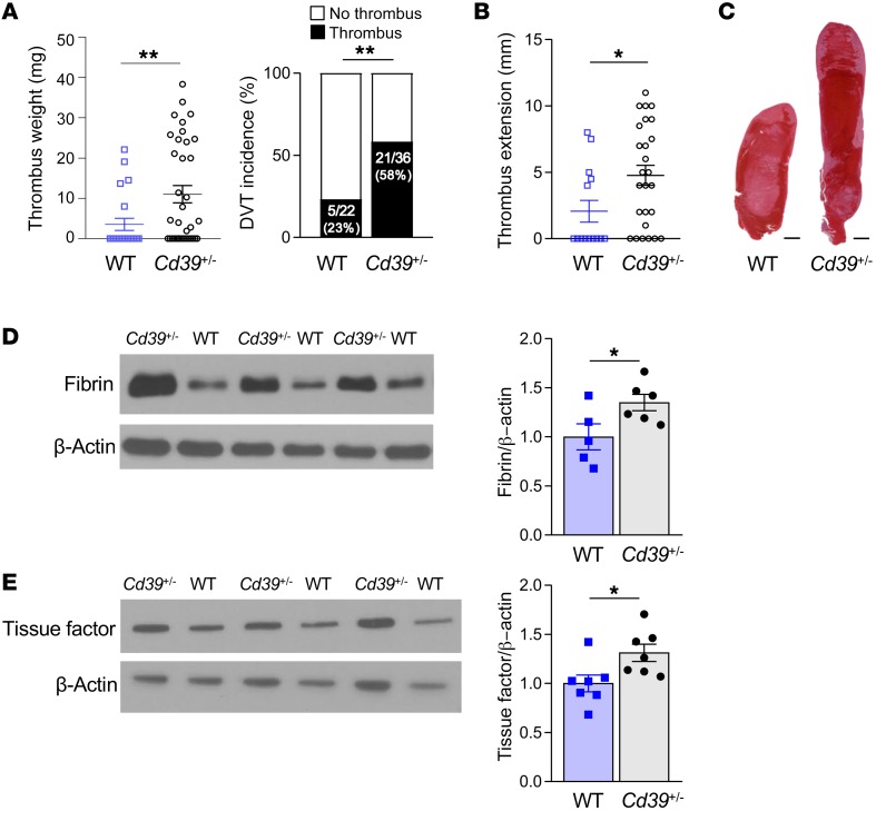
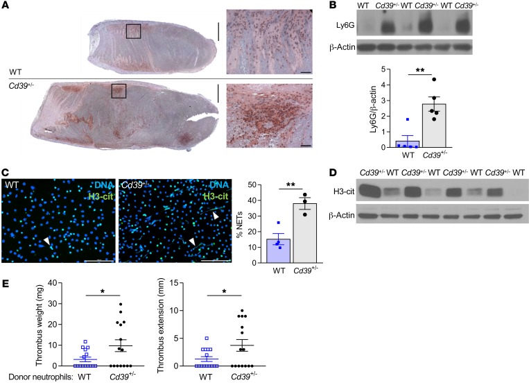
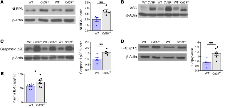
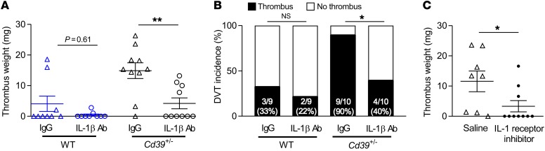
Deep vein thrombosis (DVT), caused by alterations in venous homeostasis is the third most common cause of cardiovascular mortality; however, key molecular determinants in venous thrombosis have not been fully elucidated. Several lines of evidence indicate that DVT occurs at the intersection of dysregulated inflammation and coagulation. The enzyme ectonucleoside tri(di)phosphohydrolase (ENTPD1, also known as CD39) is a vascular ecto-apyrase on the surface of leukocytes and the endothelium that inhibits intravascular inflammation and thrombosis by hydrolysis of phosphodiester bonds from nucleotides released by activated cells. Here, we evaluated the contribution of CD39 to venous thrombosis in a restricted-flow model of murine inferior vena cava stenosis. CD39-deficiency conferred a >2-fold increase in venous thrombogenesis, characterized by increased leukocyte engagement, neutrophil extracellular trap formation, fibrin, and local activation of tissue factor in the thrombotic milieu. This was orchestrated by increased phosphorylation of the p65 subunit of NFκB, activation of the NLRP3 inflammasome, and interleukin-1β (IL-1β) release in CD39-deficient mice. Substantiating these findings, an IL-1β-neutralizing antibody attenuated the thrombosis risk in CD39-deficient mice. These data demonstrate that IL-1β is a key accelerant of venous thrombo-inflammation, which can be suppressed by CD39. CD39 inhibits in vivo crosstalk between inflammation and coagulation pathways, and is a critical vascular checkpoint in venous thrombosis.

Keywords: Cardiovascular disease; Inflammation; Innate immunity; Thrombosis; Vascular Biology.


Conflict of interest statement

Conflict of interest: YK and DJP are co-inventors on an unrelated patent (application no. 20180369278). DJP is an inventor on unrelated patents (patent application no. 2017192553A1; and US patent nos. 9789133, held by Proterris Inc., 8128963, held by Columbia University, and 6972175). MH is an inventor on unrelated patents (US patent nos. 10266488, 9750757, and 9789087B2; and patent application nos. PCT/US17/38994, PCT/US19/27785, and PCT/US19/27881).

Figures

Figure 1
Figure 1. Increased venous thrombogenesis, clot extension, and fibrin content in Cd39+/– mice compared with genotype controls.
(A) Thrombus weights and thrombus frequency 2 days after IVC flow restriction (stenosis) (WT n = 22, Cd39+/– n = 36). (B) Thrombus extension (WT n = 14, Cd39+/– n = 26). (C) Representative H&E-stained sections of thrombi (n = 5, each). Scale bars: 1 mm. (D) Immunoblots of fibrin content of thrombus lysates (WT n = 5, Cd39+/– n = 6) and (E) tissue factor (n = 7 for each). Data represent the mean ± SEM. *P < 0.05 and **P < 0.01, by 2-tailed Student’s t test (D and E) with Welch’s correction (A and B). Data shown in the right panel in A were analyzed using the χ2 method.
Figure 2
Figure 2. CD39 tempers thrombus cellularity and leukocyte recruitment to growing thrombus.
(A) Neutrophil-stained (Ly6G) venous thrombi (brown, Ly6G; n = 5 for each). Scale bars: 1 mm and 50 μm (insets). (B) Immunoblot for Ly6G in venous thrombi from WT and Cd39+/– mice (n = 5 for each). (C) H3-cit expression indicates NET formation by immunofluorescence microscopy following ex vivo stimulation with 100 nM PMA (green, H3-cit; blue, DNA; white arrowheads, NETs). WT n = 4, and Cd39+/– n = 3. Scale bars: 100 μm. Representative images are shown. (D) H3-cit content in venous thrombus lysates (n = 4 for each). (E) Venous thrombus weight and extension in WT mice following adoptive transfer of WT or Cd39+/– neutrophils (n = 15 for each). Data represent the mean ± SEM. *P < 0.05 and **P < 0.01, by 2-tailed Student’s t test.
Figure 3
Figure 3. CD39 regulates inflammasome activation in mice with flow-restricted venous thrombosis.
Expression of (A) NLRP3 (WT n = 4, Cd39+/– n = 5), (B) ASC, (C) cleaved caspase-1 (p20 fragment) (n = 5 for each), and (D) mature IL-1β in thrombus lysates (WT n = 5, Cd39+/– n = 6). (E) plasma IL-1β expression by ELISA (WT n = 8, Cd39+/– n = 7). Data represent the mean ± SEM. *P < 0.05 and **P < 0.01, by 2-tailed Student’s t test.
Figure 4
Figure 4. IL-1β inhibition reduces the incidence of venous thrombosis in CD39-deficient mice.
(A) Thrombus size and (B) frequency of DVT in WT and Cd39+/– mice administered IL-1β–neutralizing or IgG isotype control antibody (200 μg i.p. 24 hours prior to surgery, n = 9–10 for each). (C) Thrombus size in Cd39+/– mice treated with vehicle control or the IL-1 receptor inhibitor anakinra (100 mg/kg/day i.p., n = 8–10 for each). Data represent the mean ± SEM. *P < 0.05 and **P < 0.01, by 2-tailed Student’s t test with Welch’s correction (A and C) and χ2 method (B).

References

    1. Marcus AJ, Broekman MJ, Pinsky DJ. COX inhibitors and thromboregulation. N Engl J Med. 2002;347(13):1025–1026. doi: 10.1056/NEJMcibr021805. - DOI - PubMed
    1. Kanthi YM, Sutton NR, Pinsky DJ. CD39: Interface between vascular thrombosis and inflammation. Curr Atheroscler Rep. 2014;16(7):425. doi: 10.1007/s11883-014-0425-1. - DOI - PMC - PubMed
    1. Hyman MC, et al. Self-regulation of inflammatory cell trafficking in mice by the leukocyte surface apyrase CD39. J Clin Invest. 2009;119(5):1136–1149. doi: 10.1172/JCI36433. - DOI - PMC - PubMed
    1. Kanthi Y, et al. Flow-dependent expression of ectonucleotide tri(di)phosphohydrolase-1 and suppression of atherosclerosis. J Clin Invest. 2015;125(8):3027–3036. doi: 10.1172/JCI79514. - DOI - PMC - PubMed
    1. Pinsky DJ, et al. Elucidation of the thromboregulatory role of CD39/ectoapyrase in the ischemic brain. J Clin Invest. 2002;109(8):1031–1040. doi: 10.1172/JCI10649. - DOI - PMC - PubMed

Publication types

MeSH terms