. 2019 Jul:208:32-44.
doi: 10.1016/j.biomaterials.2019.04.001. Epub 2019 Apr 4.

Injectable osteogenic microtissues containing mesenchymal stromal cells conformally fill and repair critical-size defects

Affiliations

Injectable osteogenic microtissues containing mesenchymal stromal cells conformally fill and repair critical-size defects

Ramkumar T Annamalai et al. Biomaterials. 2019 Jul.

Abstract

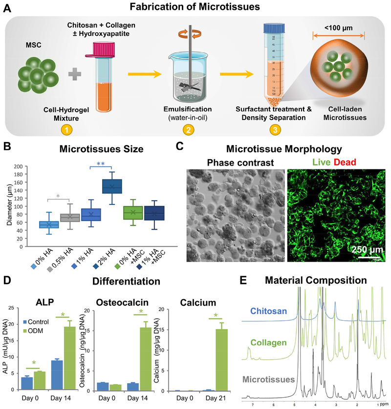
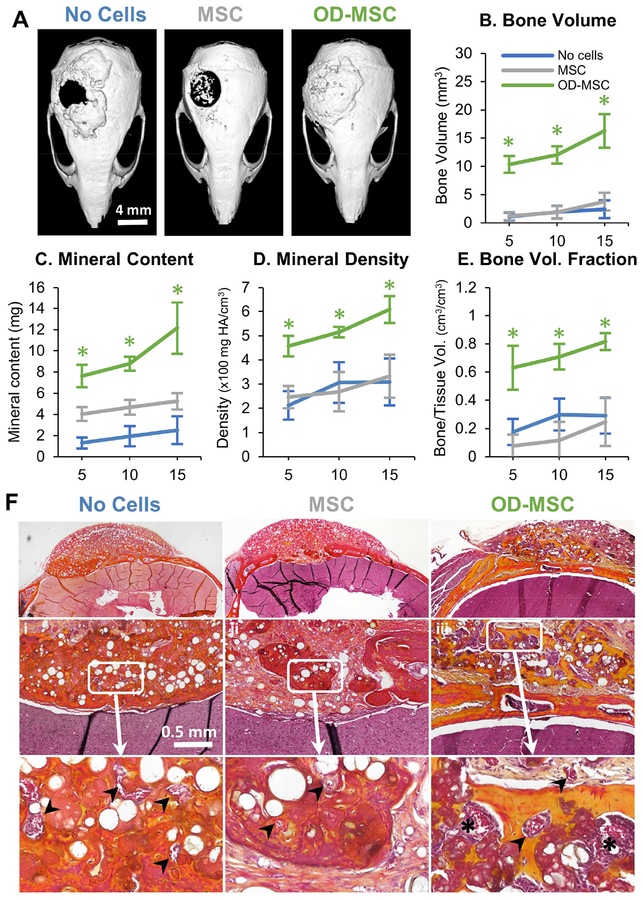
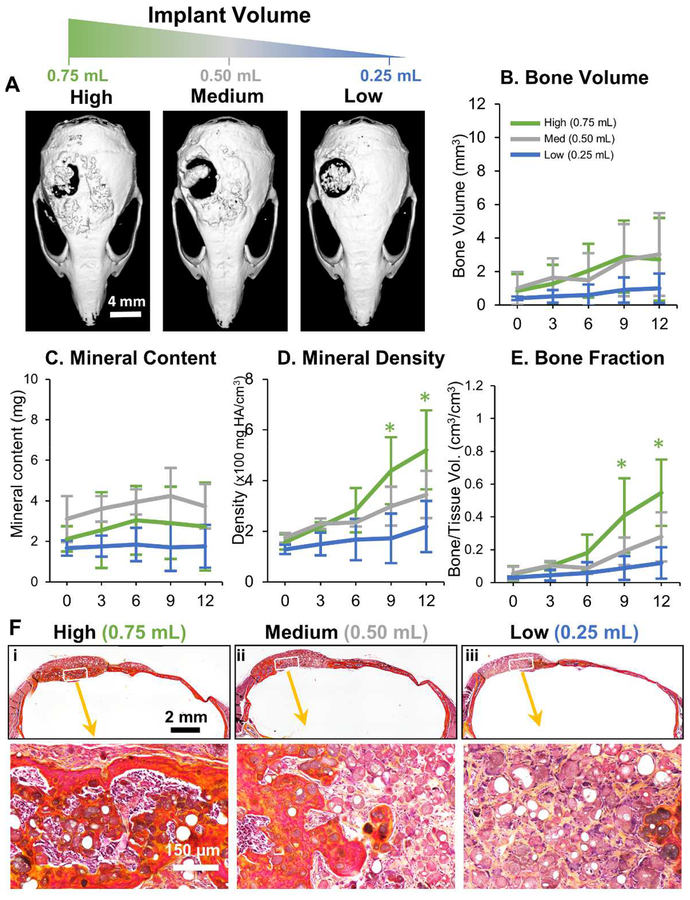
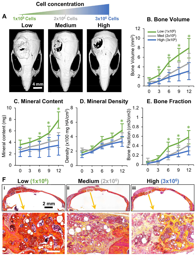
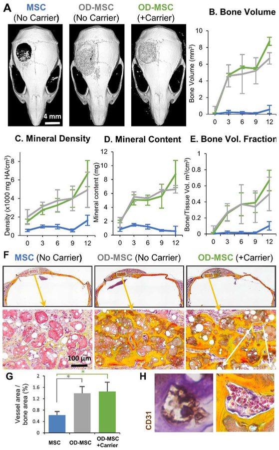
Repair of complex fractures with bone loss requires a potent, space-filling intervention to promote regeneration of bone. We present a biomaterials-based strategy combining mesenchymal stromal cells (MSC) with a chitosan-collagen matrix to form modular microtissues designed for delivery through a needle to conformally fill cavital defects. Implantation of microtissues into a calvarial defect in the mouse showed that osteogenically pre-differentiated MSC resulted in complete bridging of the cavity, while undifferentiated MSC produced mineralized tissue only in apposition to native bone. Decreasing the implant volume reduced bone regeneration, while increasing the MSC concentration also attenuated bone formation, suggesting that the cell-matrix ratio is important in achieving a robust response. Conformal filling of the defect with microtissues in a carrier gel resulted in complete healing. Taken together, these results show that modular microtissues can be used to augment the differentiated function of MSC and provide an extracellular environment that potentiates bone repair.

Keywords: Bone regeneration; Chitosan and collagen; Critical size defect; Mesenchymal stromal cells; Microtissues; Non-invasive delivery.


Figures

Figure 1 -
Figure 1 -. Fabrication and characterization of chitosan-collagen microtissues.
A) Schematic of the water-in-oil emulsification process used to embed MSC within CHI-COL composite microtissues. B) Size distribution of CHI-COL microtissues as a function of HA content and cell loading. C) Phase contrast (Day 0) and fluorescence images (Day 14) showing the morphology of microtissues and vital staining (green) of embedded MSC, respectively. D) Expression of osteogenic markers by microtissues cultured in control and osteogenic medium (ODM) in vitro. E) 1H-NMR spectra of the microtissue matrix materials, demonstrating the presence of collagen and chitosan.
Figure 2 -
Figure 2 -. Bone regeneration in a critical-sized calvarial defect.
A) Representative microCT images of bone formation in the defect region at 10 weeks. MicroCT data were analyzed to specifically assess new bone in the 4 mm defect site across implant replicates, and to obtain quantitative measures of: B) total bone volume, C) mineral content, D) mineral density, and E) bone volume fraction (bone volume/tissue volume). F) Histology images of newly formed bone in the defect site using Movat’s pentachrome staining. (Collagen fibers= yellow; fibrin = bright red; nuclei = purple-black). Arrows indicate microvessels and * indicates primitive marrow cavities.
Figure 3 -
Figure 3 -. Influence of microtissue implant volume on bone formation.
A) Representative microCT images of bone formation in the defect region at 12 weeks. MicroCT data were analyzed to specifically assess new bone within the 4 mm defect site across implant replicates, and to obtain quantitative measures of: B) total bone volume, C) mineral content, D) mineral density, and E) bone volume fraction (bone volume/tissue volume). F) Histology images of newly formed bone in the defect site using Movat’s pentachrome staining. (Collagen fibers = yellow; fibrin = bright red; nuclei = purple-black).
Figure 4 -
Figure 4 -. Influence of cell concentration on bone formation.
A) Representative microCT images of bone formation in the defect region at 12 weeks. MicroCT data were analyzed to specifically assess new bone within the 4 mm defect site across implant replicates, and to obtain quantitative measures of B) total bone volume, C) mineral content, D) mineral density, and E) bone volume fraction (bone volume/tissue volume). F) Histology images of newly formed bone in the defect site using Movat’s pentachrome staining. (Collagen fibers = yellow; fibrin = bright red; nuclei = purple-black).
Figure 5 -
Figure 5 -. Effect of microtissue delivery within a fibrin carrier gel.
A) Representative microCT images of bone formation in the defect region at 12 weeks. MicroCT data were analyzed to specifically assess new bone within the 4 mm defect site across implant replicates, and to obtain quantitative measures of B) total bone volume, C) mineral content, D) mineral density, and E) bone volume fraction (bone volume/tissue volume). F) Histology images of newly formed bone in the defect site using Movat’s pentachrome staining. (Collagen fibers = yellow; fibrin = bright red; nuclei = purple-black). G) Blood vessel area within the defect was quantified from H) histology images, which showed well-developed vessels containing erythrocytes throughout the volume of OD-MSC microtissue implants.
Figure 6 -
Figure 6 -. Ultrasound-guided, minimally invasive delivery of microtissues.
A) Schematic of the monitoring microtissue implantation into the calvarial defect using high-resolution ultrasound imaging. B) 3D ultrasound image reconstructions showing the injection of microtissues through the skin into the calvarial defect (series i-iv). C) Correlation of mineral content of microtissues and the midband fit parameter generated by spectral ultrasound imaging (SUSI). D) Heat maps of acoustic concentration generated by SUSI, showing spatial distribution and concentration of mineral in the microtissue implants.

References

    1. Henkel J, et al. , Bone Regeneration Based on Tissue Engineering Conceptions - A 21st Century Perspective. Bone Res, 2013. 1(3): p. 216–48. - PMC - PubMed
    1. Dimitriou R, et al. , Complications following autologous bone graft harvesting from the iliac crest and using the RIA: A systematic review. Injury, 2011. 42(Supplement 2): p. S3–S15. - PubMed
    1. Arrington ED, et al. , Complications of iliac crest bone graft harvesting. Clin Orthop Relat Res, 1996(329): p. 300–9. - PubMed
    1. Janicki P and Schmidmaier G, What should be the characteristics of the ideal bone graft substitute? Combining scaffolds with growth factors and/or stem cells. Injury, 2011. 42(Supplement 2): p. S77–S81. - PubMed
    1. Wang S, et al. , Maintenance of phenotype and function of cryopreserved bone-derived cells. Biomaterials, 2011. 32(15): p. 3739–49. - PubMed

Publication types