Review
. 2019:22:101788.
doi: 10.1016/j.nicl.2019.101788. Epub 2019 Mar 26.

How pattern information analyses of semantic brain activity elicited in language comprehension could contribute to the early identification of Alzheimer's Disease

Affiliations
Review

How pattern information analyses of semantic brain activity elicited in language comprehension could contribute to the early identification of Alzheimer's Disease

Andrew James Anderson et al. Neuroimage Clin. 2019.

Abstract

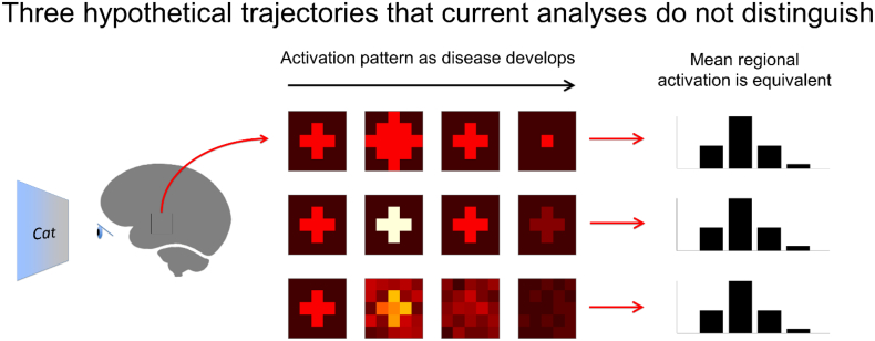
Alzheimer's disease (AD) is associated with a loss of semantic knowledge reflecting brain pathophysiology that begins years before dementia. Identifying early signs of pathophysiology induced dysfunction in the neural systems that access and process words' meaning could therefore help forecast dementia. This article reviews pioneering studies demonstrating that abnormal functional Magnetic Resonance Imaging (fMRI) response patterns elicited in semantic tasks reflect both AD-pathophysiology and the hereditary risk of AD, and also can help forecast cognitive decline. However, to bring current semantic task-based fMRI research up to date with new AD research guidelines the relationship with different types of AD-pathophysiology needs to be more thoroughly examined. We shall argue that new analytic techniques and experimental paradigms will be critical for this. Previous work has relied on specialized tests of specific components of semantic knowledge/processing (e.g. famous name recognition) to reveal coarse AD-related changes in activation across broad brain regions. Recent computational advances now enable more detailed tests of the semantic information that is represented within brain regions during more natural language comprehension. These new methods stand to more directly index how pathophysiology alters neural information processing, whilst using language comprehension as the basis for a more comprehensive examination of semantic brain function. We here connect the semantic pattern information analysis literature up with AD research to raise awareness to potential cross-disciplinary research opportunities.


Figures

Fig. 1
Fig. 1
Pyramids and Palm Trees example stimuli and results from Adamczuk et al. (2016). (Top) “Stimuli and tasks in fMRI experiment. Associative-semantic task with words (blue) and with pictures (purple). Visuoperceptual task with words (cyan) or pictures (yellow). Resting baseline with fixation point (red). Subjects were asked to press a left- or right-hand key depending on which of the 2 lower stimuli matched the upper stimulus more closely in meaning (blue, purple) or in size on the screen (cyan, yellow). A given concept triplet was presented in either the word or the picture format, and this was counterbalanced across subjects. Arrow in the top of the figure shows a timeline of 1 fMRI run, with each condition indicated in its respective color. The order of conditions was randomized for each run and subject. Translation: deur = door, hek = fence, raam = window.” (Bottom) “Area in the left posterior MTG of significant correlation between amyloidosis (SUVRcomp) and fMRI response during associative-semantic minus visuoperceptual condition (Contrast 1) (cluster peak −57, −45, 9, ext = 64 voxels, cluster-level Pcorrected = 0.006). The color scale indicates the T-values. MNI coordinates are indicated in the left upper corner and orientation of the brain in the right upper corner.” Figures reproduced with permission. We note here that whilst the visuoperceptual condition controls for the visual appearance of word/picture stimuli, it is likely to have placed lower demands on working memory. This is because unlike the associative-semantic condition it did not require the meaning of three words to be stored in working memory and compared). Consequently, the contrast map (bottom) may partially reflect this.
Fig. 2
Fig. 2
a. From Woodard et al. (2009). “Regions (shown in blue) demonstrating significant differences between the Famous and Unfamiliar Name conditions, conducted separately for each of the three groups. Brain activation projected on the lateral and medial surfaces of the left and right hemispheres.” Figure reproduced with permission. The b annotation is newly inserted in the current article to facilitate comparison with Fig. 3, Fig. 4.
Fig. 3
Fig. 3
From Seidenberg et al. (2009). “Results of voxel-wise analysis demonstrating significant differences between the famous and unfamiliar name conditions, conducted separately for each group: control (CON), family history (FH), and family history and APOEε4 (FH + ε4) groups. Yellow = regions showing greater activation to famous than unfamiliar names; blue = regions showing greater activation to unfamiliar than famous names. Brain activation projected on the lateral and medial surfaces of the left and right hemispheres.” Figure reproduced with permission.
Fig. 4
Fig. 4
From Rao et al. (2015). “Voxelwise subtraction of the Famous and Non-Famous Name hemodynamic response functions for the Low Risk and APOE ε4 groups at baseline (0 months), 18 months, and 57 months.” Figure reproduced with permission.
Fig. 5
Fig. 5
Left. Difference in Amyloid and Tau accumulation and neurodegeneration in 30 amyloid PET-positive patients with mild probable AD comparative 12 amyloid PET-negative healthy controls (Iaccarino et al., 2018). Right. The semantic network as identified and interpreted by Binder and Desai (2011).
Fig. 6
Fig. 6
How current metrics of whole region activation could overlook changes in information within brain regions.
Fig. 7
Fig. 7
A simple computational text-based semantic model of word meaning.
Fig. 8
Fig. 8
Representational similarity analysis (RSA), indexing the semantic information content in a brain region using a semantic model (e.g. Fig. 7).
Fig. 9
Fig. 9
Predicting fMRI activation elicited whilst listening to natural speech using acoustic, grammatical and semantic features (left). Predicting acoustic, grammatical and semantic features from fMRI activation (right).

Similar articles

Cited by

References

    1. Adamczuk K., De Weer A.S., Nelissen N., Dupont P., Sunaert S., Bettens K., Sleegers K., Van Broeckhoven C., Van Laere K., Vandenberghe R. Functional changes in the language network in response to increased amyloid beta deposition in cognitively intact older adults. Cereb. Cortex. 2016;26:358–373. - PubMed
    1. Ahmed S., Arnold R., Thompson S.A., Graham K.S., Hodges J.R. Naming of objects, faces and buildings in mild cognitive impairment. Cortex. 2008;44:746–752. - PubMed
    1. Albert M.S., DeKosky S.T., Dickson D., Dubois B., Feldman H.H., Fox N.C., Gamst A., Holtzman D.M., Jagust W.J., Petersen R.C., Snyder P.J., Carrillo M.C., Thies B., Phelps C.H. The diagnosis of mild cognitive impairment due to Alzheimer's disease: recommendations from the National Institute on Aging-Alzheimer's Association workgroups on diagnostic guidelines for Alzheimer's disease. Alzheimers Dement. 2011;7:270–279. - PMC - PubMed
    1. Anderson A.J., Bruni E., Bordignon U., Poesio M., Baroni M. Proceedings of the 2013 Conference on Empirical Methods in Natural Language Processing. 2013. Of words, eyes and brains: Correlating image-based distributional semantic models with neural representations of concepts; pp. 1960–1970.
    1. Anderson A.J., Bruni E., Lopopolo A., Poesio M., Baroni M. Reading visually embodied meaning from the brain: visually grounded computational models decode visual-object mental imagery induced by written text. Neuroimage. 2015;120:309–322. - PubMed

Publication types