18F-FDG-PET Can Predict Microvessel Density in Head and Neck Squamous Cell Carcinoma
- PMID: 30991696
- PMCID: PMC6521262
- DOI: 10.3390/cancers11040543
18F-FDG-PET Can Predict Microvessel Density in Head and Neck Squamous Cell Carcinoma
Abstract
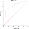
Aim: Positron emission tomography (PET) with 18F-fluordeoxyglucose (18F-FDG) plays an essential role in the staging and tumor monitoring of head and neck squamous cell carcinoma (HNSCC). Microvessel density (MVD) is one of the clinically important histopathological features in HNSCC. The purpose of this study was to analyze possible associations between 18F-FDG-PET findings and MVD parameters in HNSCC. Materials and Methods: Overall, 22 patients with a mean age of 55.2 ± 11.0 and with different HNSCC were acquired. In all cases, whole-body 18F-FDG-PET was performed. For each tumor, the maximum and mean standardized uptake values (SUVmax; SUVmean) were determined. The MVD, including stained vessel area and total number of vessels, was estimated on CD105 stained specimens. All specimens were digitalized and analyzed by using ImageJ software 1.48v. Spearman's correlation coefficient (r) was used to analyze associations between investigated parameters. p-values of <0.05 were taken to indicate statistical significance. Results: SUVmax correlated with vessel area (r = 0.532, p = 0.011) and vessel count (r = 0.434, p = 0.043). Receiver operating characteristic analysis identified a threshold SUVmax of 15 to predict tumors with high MVD with a sensitivity of 72.7% and specificity of 81.8%, with an area under the curve of 82.6%. Conclusion: ⁸F-FDG-PET parameters correlate statistically significantly with MVD in HNSCC. SUVmax may be used for discrimination of tumors with high tumor-related MVD.
Keywords: head and neck neoplasms; neovascularization; pathologic; positron emission tomography.
Conflict of interest statement
The authors declare no conflict of interest.
Figures


Similar articles
-
Associations Between [18F]FDG-PET and Complex Histopathological Parameters Including Tumor Cell Count and Expression of KI 67, EGFR, VEGF, HIF-1α, and p53 in Head and Neck Squamous Cell Carcinoma.Mol Imaging Biol. 2019 Apr;21(2):368-374. doi: 10.1007/s11307-018-1223-x. Mol Imaging Biol. 2019. PMID: 29931433
-
Simultaneous (18)F-FDG-PET/MRI: Associations between diffusion, glucose metabolism and histopathological parameters in patients with head and neck squamous cell carcinoma.Oral Oncol. 2016 Jul;58:14-20. doi: 10.1016/j.oraloncology.2016.04.009. Epub 2016 May 7. Oral Oncol. 2016. PMID: 27311397
-
Parameters of simultaneous 18F-FDG-PET/MRI predict tumor stage and several histopathological features in uterine cervical cancer.Oncotarget. 2017 Apr 25;8(17):28285-28296. doi: 10.18632/oncotarget.16043. Oncotarget. 2017. PMID: 28423698 Free PMC article.
-
Hyperaccumulation of (18)F-FDG in order to differentiate solid pseudopapillary tumors from adenocarcinomas and from neuroendocrine pancreatic tumors and review of the literature.Hell J Nucl Med. 2013 May-Aug;16(2):97-102. doi: 10.1967/s002449910084. Epub 2013 May 20. Hell J Nucl Med. 2013. PMID: 23687644 Review.
-
Diagnostic accuracy of F-18 FDG PET or PET/CT for detection of lymph node metastasis in clinically node negative head and neck cancer patients; A systematic review and meta-analysis.Am J Otolaryngol. 2019 Mar-Apr;40(2):297-305. doi: 10.1016/j.amjoto.2018.10.013. Epub 2018 Oct 23. Am J Otolaryngol. 2019. PMID: 30473166
Cited by
-
Relationships Between DCE-MRI, DWI, and 18F-FDG PET/CT Parameters with Tumor Grade and Stage in Patients with Head and Neck Squamous Cell Carcinoma.Mol Imaging Radionucl Ther. 2021 Oct 15;30(3):177-186. doi: 10.4274/mirt.galenos.2021.25633. Mol Imaging Radionucl Ther. 2021. PMID: 34658826 Free PMC article.
-
Correlation between histogram-based DCE-MRI parameters and 18F-FDG PET values in oropharyngeal squamous cell carcinoma: Evaluation in primary tumors and metastatic nodes.PLoS One. 2020 Mar 2;15(3):e0229611. doi: 10.1371/journal.pone.0229611. eCollection 2020. PLoS One. 2020. PMID: 32119697 Free PMC article.
-
What Is the Role of Imaging in Cancers?Cancers (Basel). 2020 Jun 8;12(6):1494. doi: 10.3390/cancers12061494. Cancers (Basel). 2020. PMID: 32521685 Free PMC article.
References
-
- Varoquaux A., Rager O., Poncet A., Delattre B.M., Ratib O., Becker C.D., Dulguerov P., Dulguerov N., Zaidi H., Becker M. Detection and quantification of focal uptake in head and neck tumours: (18)F-FDG PET/MR versus PET/CT. Eur. J. Nucl. Med. Mol. Imaging. 2014;41:462–475. doi: 10.1007/s00259-013-2580-y. - DOI - PMC - PubMed
-
- Haerle S.K., Huber G.F., Hany T.F., Ahmad N., Schmid D.T. Is there a correlation between 18F-FDG-PET standardized uptake value, T-classification, histological grading and the anatomic subsites in newly diagnosed squamous cell carcinoma of the head and neck? Eur. Arch. Otorhinolaryngol. 2010;267:1635–1640. doi: 10.1007/s00405-010-1348-2. - DOI - PubMed
-
- Li S.J., Guo W., Ren G.X., Huang G., Chen T., Song S.L. Expression of Glut-1 in primary and recurrent head and neck squamous cell carcinomas, and compared with 2-[18F]fluoro-2-deoxy-D-glucose accumulation in positron emission tomography. Br. J. Oral Maxillofac. Surg. 2008;46:180–186. doi: 10.1016/j.bjoms.2007.11.003. - DOI - PubMed
-
- Abgral R., Keromnes N., Robin P., Le Roux P.Y., Bourhis D., Palard X., Rousset J., Valette G., Marianowski R., Salaün P.Y. Prognostic value of volumetric parameters measured by 18F-FDG PET/CT in patients with head and neck squamous cell carcinoma. Eur. J. Nucl. Med. Mol. Imaging. 2014;41:659–667. doi: 10.1007/s00259-013-2618-1. - DOI - PubMed
LinkOut - more resources
Full Text Sources

