. 2019 Jul;33(7):8386-8405.
doi: 10.1096/fj.201802281R. Epub 2019 Apr 16.

Distinct expression pattern of periostin splice variants in chondrocytes and ligament progenitor cells

Affiliations

Distinct expression pattern of periostin splice variants in chondrocytes and ligament progenitor cells

Lei Cai et al. FASEB J. 2019 Jul.

Abstract

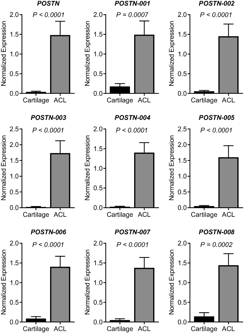
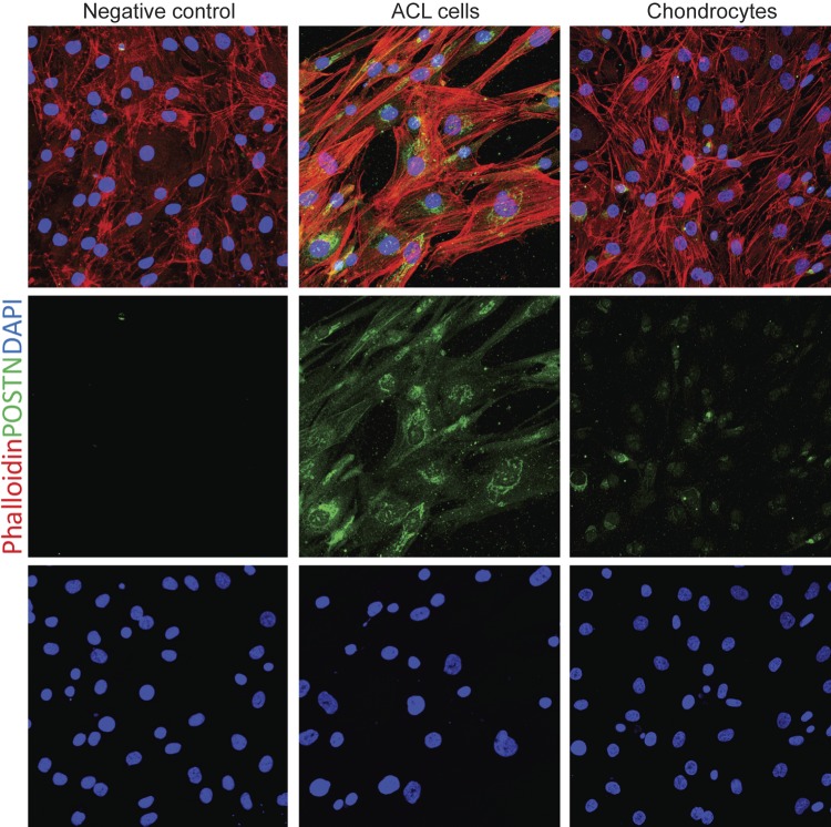
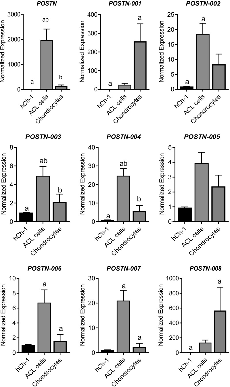
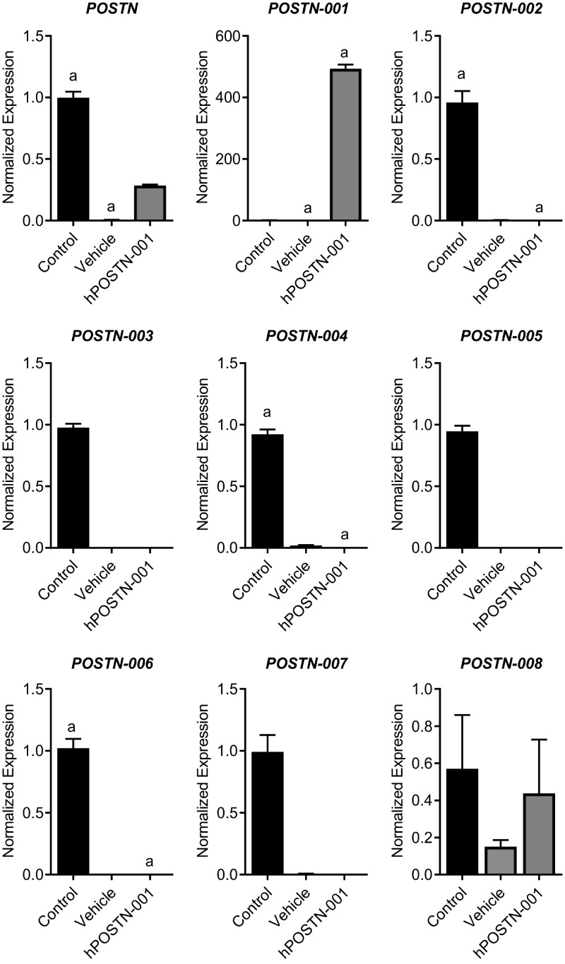
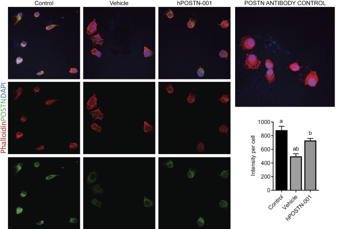
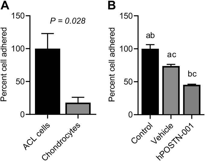
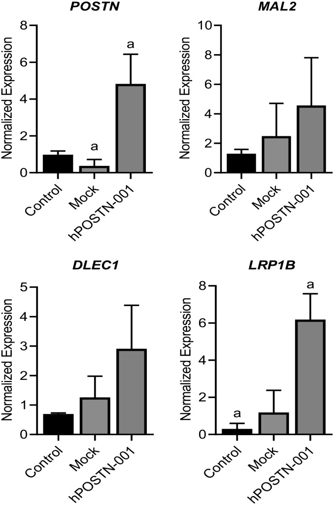
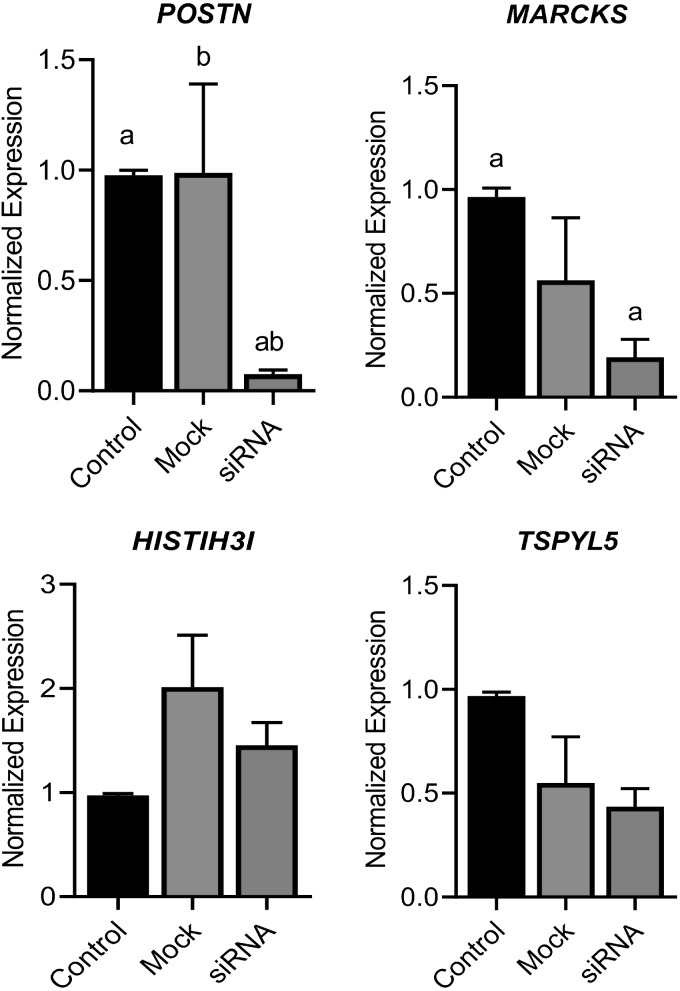
Periostin (POSTN), a secretory matricellular matrix protein, plays a multitude of biologic functions. Various splice variants of POSTN have been described; however, their expression pattern and functional implications are not completely understood. This study was undertaken to decipher the differential expression pattern of POSTN and its splice variants in various tissues and cell types. We show that POSTN was more highly expressed in anterior cruciate ligament (ACL) remnants compared with articular cartilage at the cellular and tissue level. Isoforms 1 and 8 were highly expressed only in articular chondrocytes, suggesting their splice-specific regulation in chondrocytes. To discern the role of total POSTN and full-length human POSTN isoform 1 (hPOSTN-001), we stably transfected human chondrosarcoma 1 (hCh-1) cell line with hPOSTN-001 using a pcDNA3.1-hPOSTN-001 construct. RNA-sequencing analysis of hCh-1 cells identified differentially expressed genes with a known role in chondrocyte function and osteoarthritis. Similar expression of a subset of candidate genes was revealed in ACL progenitor cells and chondrocytes as well as in ACL progenitor cells in which POSTN activity was altered by overexpression and by small interfering RNA gene knockdown. Cells expressing total POSTN, not isoform 1, exhibited increased cell adhesion potential. These findings suggest an important role for POSTN in the knee.-Cai, L., Brophy, R. H., Tycksen, E. D., Duan, X., Nunley, R. M., Rai, M. F. Distinct expression pattern of periostin splice variants in chondrocytes and ligament progenitor cells.

Keywords: RNA-seq; anterior cruciate ligament; cartilage; osteoarthritis; periostin isoforms.


Conflict of interest statement

The authors thank Washington University Genome Technology Access Center (GTAC) for assistance with RNA-seq analysis. This study was supported by research funds provided by the Department of Orthopaedic Surgery, Washington University. Dr. M. F. Rai is supported through the Pathway to Independence Award (R00-AR-064837) from the U.S. National Institutes of Health (NIH), National Institute of Arthritis and Musculoskeletal and Skin Diseases (NIAMS). The content of this publication is solely the responsibility of the authors and does not necessarily represent the official views of the NIH or NIAMS. The authors declare no conflicts of interest.

Figures

Figure 1
Figure 1
Expression pattern of POSTN and its splice variants in cartilage and ACL tissues. To determine whether POSTN and its splice variants are differentially expressed, we compared the expressions profile between cartilage (n = 8) and ACL remnants (n = 12). We observed that POSTN and all 8 transcript variants were significantly more highly expressed in ACL tissue than cartilage.
Figure 2
Figure 2
POSTN immunostaining in ACL cells and chondrocytes. To confirm and compare POSTN protein in ACL progenitor cells and chondrocytes, we performed the immunostaining for POSTN. Immunostaining of human ACL progenitor cells (n = 3) and chondrocytes (n = 3) revealed that POSTN was highly present in ACL cells, whereas chondrocytes showed faint signal for POSTN. Phalloidin (red-orange) was used to visualize F-actin, DAPI (blue) staining showed nuclei, and POSTN was shown by green staining. For POSTN antibody control, we used chondrocytes as shown in the left panel. Magnification value, ×400.
Figure 3
Figure 3
Expression pattern of POSTN and its splice variants in hCh-1 cells, chondrocytes, and ACL progenitor cells. To determine whether POSTN and its splice variants are differentially expressed, we compared the expressions profile in hCh-1 cells (n = 3), articular chondrocytes (n = 9), and ACL progenitor cells (n = 6). We observed that POSTN mRNA was significantly higher in ACL progenitor cells compared with both hCh-1 cells and chondrocytes. Expression of splice variants showed 3 patterns: those highly expressed in chondrocytes (isoforms 1 and 8), those highly expressed in ACL progenitor cells (isoforms 2, 3, 4, 6 and 7), and those that showed no significant difference (isoform 5). Similar lowercase letters (a, b) in each graph represent statistical significance from each other at P < 0.05.
Figure 4
Figure 4
Expression pattern of POSTN and its splice variants in hCh-1 cells stably transfected clones. To determine whether POSTN and its splice variants are differentially expressed, we compared the expression profile of nontransfected hCh-1 cells (control, n = 3), vehicle cells (vehicle, n = 3), and cells transfected with pcDNA3.1–hPOSTN-001 (hPOSTN-001, n = 3). We observed that POSTN mRNA was significantly higher in control compared with vehicle cells. POSTN isoform 1 was significantly higher on hPOSTN-001 cells compared with control and vehicle cells. Lowercase letters (a) in each graph represent statistical significance from each other at P < 0.05.
Figure 5
Figure 5
POSTN immunostaining in hCh-1 cells. To detect POSTN protein in hCh-1 nontransfected cells (control, n = 3), vehicle-transfected cells (vehicle, n = 3), and cells transfected with pcDNA3.1-hPOSTN-001 (hPOSTN-001, n = 3), cells were subjected to immunofluorescence. Phalloidin (red-orange) was used to visualize F-actin, DAPI (blue) staining showed nuclei, and POSTN was shown by green staining. Magnification value, ×400. Quantification showed that control and hPOSTN-001 cells had significantly higher levels of POSTN signals than vehicle cells. These data reflected the same pattern as shown by real-time PCR. Similar lowercase letters (a, b) in each graph represent statistical significance from each other at P < 0.05.
Figure 6
Figure 6
RNA-seq analysis. A) End bias plot showing the alignment of all reads across a theoretical 100-bp normalized gene body. All samples (n = 3 per group) showed excellent coverage from 3′ to 5′, indicating that sampling of exons for isoform quantification should be uniform and robust. B) Junction saturation plot illustrating the random sampling of reads from all aligned BAM files for each sample in 5% intervals, where the total number of reads spanning known exon-exon junctions are summed for each interval and plotted to find the point of junction saturation. A plateau for each sample at or near 100% sampling indicates that each sample was sequenced to near complete junction saturation and that any further sequencing would only increase sequencing depth for each detected gene rather than discover new genes not yet detected. C) Spearman correlation matrix of limma voomWithQualityWeights transformed expression values. D) Hierarchical clustering of the limma voomWithQualityWeights transformed expression values; the heatmap of model coefficients shows that all samples cluster as would be expected according to transfection status.
Figure 7
Figure 7
RNA-seq quantitative data. A) Three-dimensional principle components analysis illustrating that 96.4% of all variance can be accounted for in the first principle component and that all samples cluster by their respective transfection status. B, C) Venn diagrams showing number of differentially expressed transcripts in each comparison and the overlaps between any 2 or more comparisons. The numbers of transcripts shown in overlapping areas illustrate the number of transcripts common to 2 or more comparisons. All numbers are given using ANOVA at P < 0.05. The numbers of significantly differentially expressed transcripts at any fold change (B) or at ≥2-fold (C) are depicted in the form of Venn diagrams. DF) Volcano plot of all genes expressed at >1 count per million in all samples: hPOSTN-001 vs. vehicle (D), hPOSTN-001 vs. control (E), and control vs. vehicle (F). The observed log2 fold change is on the x axis and the unadjusted P value converted to the –log10 scale is on the y axis. All genes with Benjamini-Hochberg adjusted P values ≤0.05 are highlighted in red.
Figure 8
Figure 8
Effect of POSTN or hPOSTN-001 on cell adhesion. Effect of total POSTN or hPOSTN-001 was examined on cell adhesion by Vybrant cell adhesion assay kit. A) We found that ACL progenitor cells (n = 3) that had higher expression of total POSTN compared with chondrocytes (n = 3) showed more cell adhesion potential. B) Similarly, control hCh-1 cells (n = 3) that had higher expression of total POSTN compared with vehicle (n = 3) and hPOSTN-001 trasfected cells (n = 3) also had significantly higher cell adhesion. Similar lowercase letters (a, b, c) in each graph represent statistical significance from each other at P < 0.05.
Figure 9
Figure 9
Real-time PCR of selected genes in ACL progenitor cells and chondrocytes. To get some insights into the role of total POSTN and POSTN isoform 1 on selected genes from RNA-seq analysis, we found that MARCKS, HIST1H3I, and PCDHB5 were more highly expressed in ACL progenitor cells (n = 5) than in chondrocytes (n = 6) and that MAL2, DLEC1, and LRP1B were significantly more highly expressed in chondrocytes than in ACL progenitor cells. Lowercase letters (a) in each graph represent statistical significance from each other at P < 0.05.
Figure 10
Figure 10
Effect of hPOSTN-001 overexpression on selected genes in ACL progenitor cells. To validate if the effect of hPOSTN-001 on 3 genes identified from RNA-seq analysis, we performed overexpression of POSTN isoform 1 in ACL progenitor cells (n = 3). We observed that overexpression of hPOSTN-001 in ACL progenitor cells resulted in up-regulation of mRNA expression of 3 genes, namely, MAL2, DLEC1, and LRP1B. Lowercase letters (a) in each graph represent statistical significance from each other at P < 0.05.
Figure 11
Figure 11
Effect of POSTN ablation on selected genes. To validate if the effect of POSTN on 3 genes identified from RNA-seq analysis, we performed POSTN knockdown in ACL progenitor cells (n = 5). Our data showed that knockdown of total POSTN from ACL progenitor cells resulted in suppression of these 3 genes: MARCKS, HIST1H3I, and TSPYL5. Similar lowercase letters (a, b) in each graph represent statistical significance from each other at P < 0.05.

References

    1. Coutu D. L., Wu J. H., Monette A., Rivard G. E., Blostein M. D., Galipeau J. (2008) Periostin, a member of a novel family of vitamin K-dependent proteins, is expressed by mesenchymal stromal cells. J. Biol. Chem. 283, 17991–18001 - PubMed
    1. Takeshita S., Kikuno R., Tezuka K., Amann E. (1993) Osteoblast-specific factor 2: cloning of a putative bone adhesion protein with homology with the insect protein fasciclin I. Biochem. J. 294, 271–278 - PMC - PubMed
    1. Litvin J., Selim A. H., Montgomery M. O., Lehmann K., Rico M. C., Devlin H., Bednarik D. P., Safadi F. F. (2004) Expression and function of periostin-isoforms in bone. J. Cell. Biochem. 92, 1044–1061 - PubMed
    1. Kim C. J., Isono T., Tambe Y., Chano T., Okabe H., Okada Y., Inoue H. (2008) Role of alternative splicing of periostin in human bladder carcinogenesis. Int. J. Oncol. 32, 161–169 - PubMed
    1. Horiuchi K., Amizuka N., Takeshita S., Takamatsu H., Katsuura M., Ozawa H., Toyama Y., Bonewald L. F., Kudo A. (1999) Identification and characterization of a novel protein, periostin, with restricted expression to periosteum and periodontal ligament and increased expression by transforming growth factor beta. J. Bone Miner. Res. 14, 1239–1249 - PubMed

Publication types