

Effects of tranexamic acid on the recovery of osteochondral defects treated by microfracture and acellular matrix scaffold: an experimental study
- PMID: 30992060
- PMCID: PMC6469115
- DOI: 10.1186/s13018-019-1144-7
Effects of tranexamic acid on the recovery of osteochondral defects treated by microfracture and acellular matrix scaffold: an experimental study
Abstract
Background: Microfracture and scaffold application in the treatment of osteochondral defects is still one of the most frequently used methods in the clinic. The most important step in this treatment method is the stabilization of fibrin clot. Tranexamic acid (TA) is an antifibrinolytic agent commonly used in orthopedic surgery in recent years. This study evaluated the effect of local TA application on healing of experimentally induced osteochondral defects on rabbits.
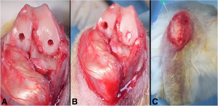
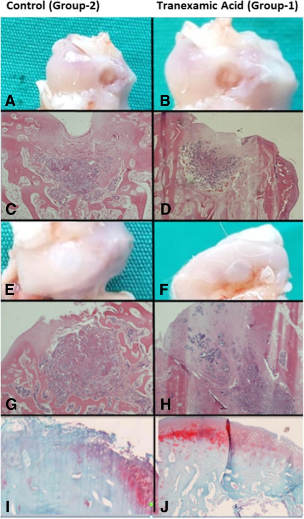
Methods: This paper contains an animal in vivo data and histological outcomes on the effect of TA. Eighteen New Zealand white rabbits were treated unilaterally and cylindrical defects having a width of 4 mm and depth of 5 mm were created in the weight-bearing surfaces of the medial and lateral condyles of the right femur. They were divided into two groups, as group 1 study and group 2 control groups, respectively. One milliliter (ml) of TA was injected into the knee joints of the subjects in group 1. All animals were sacrificed for the extraction of the femur condyles for histologic study at the fourth and eighth weeks after surgery. Histological evaluations were performed by Brittberg and O'Driscoll scores to all samples. Data were organized in a Standard Statistical Package System v.22 software package (SPSS/PC Inc., Chicago, IL.) and reported as mean and median (min-max). Repeated measures ANOVA test was used to compare groups and condyle effects together for each week. p values below 0.05 were considered as statistically significant.
Results: Samples were taken in the fourth and eighth weeks. The regularity of the surface in group 1 was smoother, and the tissue stability was more robust. Mean Brittberg scores in both weeks were statistically higher in group 1 when compared with group 2. In the microscopic evaluation, it was observed that the regeneration of subchondral and cartilage tissues were more rapid and organized in group 1, and the mean O' Driscoll scores in both weeks were statistically higher in group 1.
Conclusions: Application of TA improves the healing time and tissue stability in osteochondral defects which are implanted a-cellular scaffold after microfracture and should be applicable to humans for the treatment of osteochondral defects.
Keywords: A-cellular scaffold; Microfracture; Osteochondral defect; Tranexamic acid.
Conflict of interest statement
Ethics approval and consent to participate
This study was carried out with the approval of the local ethics committee of animal research of Abant Izzet Baysal University with the decision numbered 08.03.2017/11.
Consent for publication
Not applicable
Competing interests
The authors declare that they have no competing interests.
Publisher’s Note
Springer Nature remains neutral with regard to jurisdictional claims in published maps and institutional affiliations.
Figures
References
-
- Rastrelli A, Beccaro M, Biviano F. Hyaluronic acid esters, a new class of semisynthetic biopolymers: chemical and physico-chemical characterization. Clin Implant Mater. 1990;9:199–205.
MeSH terms
Substances
Grants and funding
LinkOut - more resources
Full Text Sources
