Review
. 2019 Jun;42(6):414-424.
doi: 10.1016/j.tins.2019.03.006. Epub 2019 Apr 13.

Mechanisms of Hair Cell Damage and Repair

Affiliations
Review

Mechanisms of Hair Cell Damage and Repair

Elizabeth L Wagner et al. Trends Neurosci. 2019 Jun.

Abstract

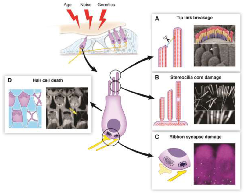
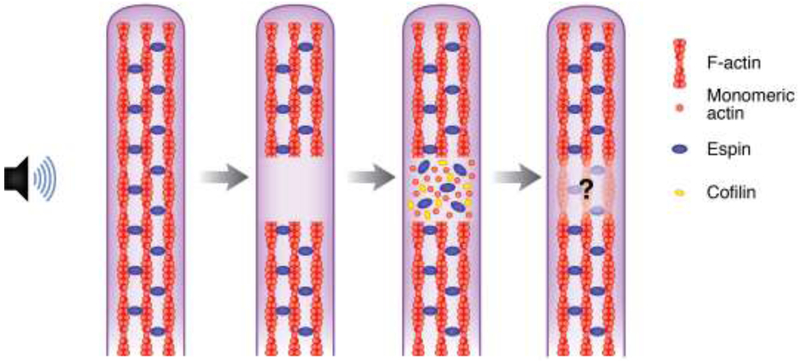
Sensory hair cells of the inner ear are exposed to continuous mechanical stress, causing damage over time. The maintenance of hair cells is further challenged by damage from a variety of other ototoxic factors, including loud noise, aging, genetic defects, and ototoxic drugs. This damage can manifest in many forms, from dysfunction of the hair cell mechanotransduction complex to loss of specialized ribbon synapses, and may even result in hair cell death. Given that mammalian hair cells do not regenerate, the repair of hair cell damage is important for continued auditory function throughout life. Here, we discuss how several key hair cell structures can be damaged, and what is known about how they are repaired.

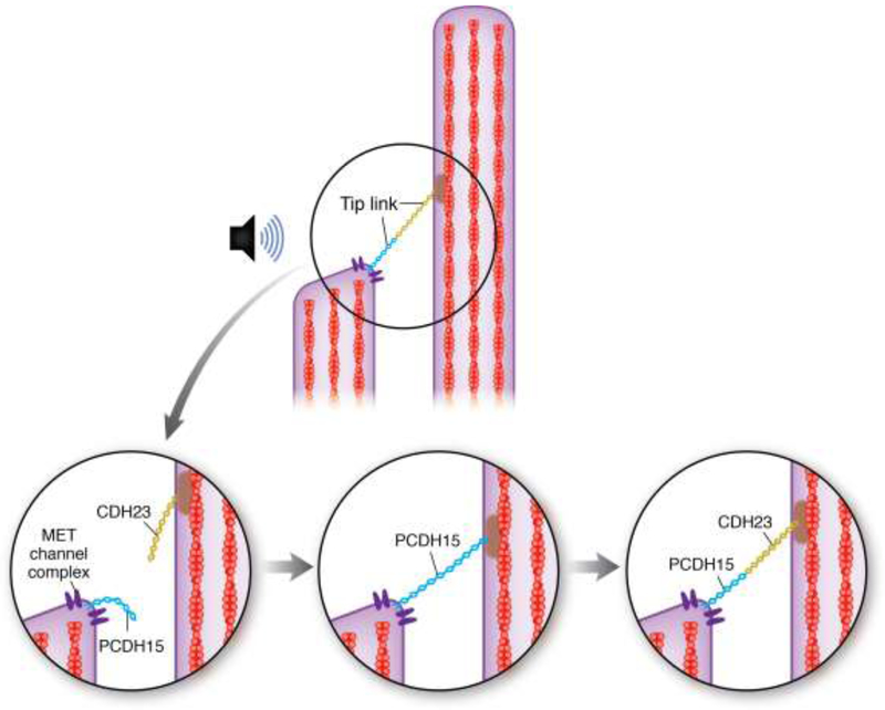
Keywords: F-actin core; Hair cell; mechanotransduction; ribbon synapse; stereocilia; tip link.


Conflict of interest statement

Competing interests

The authors declare no competing interests.

Figures

Figure 1, Key Figure.
Figure 1, Key Figure.. An Overview of Hair Cell Damage.
(Top Left) Hair cells can accumulate damage stemming from a variety of factors including age, noise, and genetics. (Center) Diagram of a hair cell indicating sites vulnerable to damage. (A) Tip link breakage. Left: Tip links can be broken by overstimulation or by in vitro calcium chelation, leading to a loss of tension on the MET channel complex and subsequent loss of the MET current. Right: Scanning electron micrograph showing an outer hair cell bundle. Tip links (white arrow) are visible at higher magnification below. (B) Stereocilia core damage. Left: Overstimulation of hair bundles causes the appearance of gaps in staining of the stereocilia F-actin core. These gaps likely represent sites of F-actin depolymerization, which would decrease bundle rigidity. Right: Phalloidin staining of a noise-damaged inner hair cell bundle. Gaps in the staining are visible at higher magnification below. (C) Ribbon synapse damage. Left: Ribbon synapses can be lost due to exposure to loud noise or prolonged exposure to milder noise, even in absence of permanent hearing threshold shift. Their loss can reduce hearing ability in noisy environments, known as “hidden hearing loss”. Right: Immunolabeling of CTBP2 (a component of ribbon synapses, green puncta) at the base of hair cells labeled by MYO7A immunostaining (magenta). (D) Hair cell death. Left: Auditory sensory epithelium with F-actin scars at the sites of missing hair cells. When hair cells die, they are extruded from the epithelium. Nearby supporting cells fill in the hole, leaving a cross-shaped F-actin scar. Right: Phalloidin labeling of F-actin in the organ of Corti from an aged mouse. An F-actin scar is seen at the site of a missing hair cell (yellow arrow). All images were taken in the Shin lab.
Figure 2.
Figure 2.. Schematic model of Tip Link Repair.
(Top) A tip link, consisting of Protocadherin 15 (blue) and Cadherin 23 (gold) connects the MET complex at the top of one stereocilium to the actin core of the stereocilium in the next tallest row. Overstimulation, such as from loud noise, can cause tip link breakage (bottom left). According to [23], a temporary tip link, consisting of only Protocadherin 15, is formed within about 12 hours of damage, partially reestablishing MET channel function (bottom middle). Within about 36 hours after damage, the Protocadherin 15 at the top half of the tip link is replaced by Cadherin 23 to allow full restoration of MET function (bottom right).
Figure 3.
Figure 3.. A Potential Mechanism for the Repair of Stereocilia F-actin Core Damage.
Overstimulation can cause the appearance of gaps in phalloidin staining of the F-actin cores of cochlear hair cell stereocilia. Gaps in F-actin staining in damaged stereocilia likely represent areas of disorganized or depolymerized actin. Immunostaining for β- and γ-actin is enriched at these sites, along with cofilin and espin. DNaseI staining is also observed, indicating that the actin is monomeric. The presence of monomeric actin, along with cofilin, which can nucleate actin at high concentrations, and espin, an actin crosslinker, suggests that localized F-actin remodeling is occurring to repair the damage [45]. However, there is no definite evidence that the damage is actually repaired.

Similar articles

Cited by

References

    1. Blackwell DL, Lucas JW,CT (2014) Summary health statistics for US adults: National Health Interview Survey. Vital Heal. Stat 10 260, 1–161 - PubMed
    1. Hudspeth AJ (2005) How the ear ‘s works work: mechanoelectrical transduction and amplification by hair cells. C.R. Biol 328, 155–162 - PubMed
    1. Fettiplace R (2017) Hair cell transduction, tuning, and synaptic transmission in the mammalian cochlea. Compr. Physiol 7, 1197–1227 - PMC - PubMed
    1. Flock A (1964) Structure of the macula utriculi with special reference to directional interplay of sensory resoneses as revealed by morphological polarization. J. Cell Biol. 22, 413–31 - PMC - PubMed
    1. Tilney LG et al. (1980) The organization of actin filaments in the stereocilia of cochlear hair cells. J. Cell Biol. 86, 244–59 - PMC - PubMed

Publication types