Review
. 2019 Apr 16;146(8):dev166074.
doi: 10.1242/dev.166074.

Brain organoids: advances, applications and challenges

Affiliations
Review

Brain organoids: advances, applications and challenges

Xuyu Qian et al. Development. .

Abstract

Brain organoids are self-assembled three-dimensional aggregates generated from pluripotent stem cells with cell types and cytoarchitectures that resemble the embryonic human brain. As such, they have emerged as novel model systems that can be used to investigate human brain development and disorders. Although brain organoids mimic many key features of early human brain development at molecular, cellular, structural and functional levels, some aspects of brain development, such as the formation of distinct cortical neuronal layers, gyrification, and the establishment of complex neuronal circuitry, are not fully recapitulated. Here, we summarize recent advances in the development of brain organoid methodologies and discuss their applications in disease modeling. In addition, we compare current organoid systems to the embryonic human brain, highlighting features that currently can and cannot be recapitulated, and discuss perspectives for advancing current brain organoid technologies to expand their applications.

Keywords: Brain organoids; Neuroscience; Stem cell.


Conflict of interest statement

Competing interestsThe authors declare no competing or financial interests.

Figures
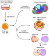
Fig. 1.
Fig. 1.
Unguided and guided approaches for making brain organoids. Unguided approaches (top) harness the intrinsic signaling and self-organization capacities of hPSCs to differentiate spontaneously into tissues mimicking the developing brain. The resulting cerebral organoids often contain heterogeneous tissues resembling various brain regions. By contrast, guided approaches (bottom) use small molecules and growth factors to generate spheroids that are specifically representative of one tissue type. Brain region-specific organoid methods involve the use of patterning factors at an early stage to specify progenitor fate; these factors are then removed in subsequent differentiation stages. Guided approaches can also be used to generate two or more spheroids/organoids representative of different brain region identities that can then be fused to form ‘assembloids’, which model interactions between different brain regions.
Fig. 2.
Fig. 2.
Structural comparison between cortical organoids and the human embryonic cortex. Cortical organoids resemble the cytoarchitecture of human developing cerebral cortex in early and mid-gestation with remarkable fidelity, despite their small size. A cortical organoid usually contains multiple short and independent neuroepithelial structures. Within each structure, well-defined layers resembling the VZ, iSVZ, oSVZ, CP and MZ can be observed. Major neural lineage cell types in the embryonic cortex can also be detected in cortical organoids, but vascular and immune cells are absent. Late-gestational features of corticogenesis, such as the formation of cortical folding and the six separated cortical layers are not observed in cortical organoids generated by currently available approaches. CP, cortical plate; IPC, intermediate progenitor cells; iSVZ, inner subventricular zone; IZ, intermediate zone; MZ, marginal zone; oRG, outer radial glia; oSVZ, outer subventricular zone; vRG, ventricular radial glia; VZ, ventricular zone.
Fig. 3.
Fig. 3.
The diffusion limit depletes progenitors and prohibits organoid expansion. Brain organoids grown as a sphere in 3D suspension cultures can expand to 3-4 mm in diameter, but only those cells within a limited distance from the surface can receive sufficient oxygen and nutrients via diffusion. The build-up of a necrotic core is therefore common in brain organoids. As the outer layers expand, the more metabolically demanding progenitors in the interior will eventually deplete, resulting in a loss of proliferation and structural disorganization over long-term cultures.
Fig. 4.
Fig. 4.
Applications of brain organoids. (1) Brain organoids have proven to be particularly informative for modeling congenital brain malformations caused by genetic deficits or infectious disease, because the organoid cytoarchitecture provide a direct read-out for disease-relevant phenotypes. (2) Primitive microcircuits are detected in brain organoids (Birey et al., 2017), but further promoting functional maturation will be key for modeling psychiatric disorders such as autism and schizophrenia. (3) It remains challenging to model age-dependent neurodegenerative diseases with current brain organoids, because they mimic mostly embryonic brain development. A method of artificially inducing aging in vitro could potentially allow organoids to represent disease-relevant phenotypes. (4) The ability to generate brain organoids in large quantities with high consistency raises the possibility of using organoids for compound screening and subsequent validation. The development of automated high-throughput platforms could expedite such advances. (5) Organoids offer the unique opportunity to understand the basis of human brain formation and evolution in comparison to other species. For instance, brain samples from great apes are largely inaccessible, but organoids generated from great ape iPSCs can be compared with human cell-derived organoids to discover uniquely human features.

References

    1. Abud E. M., Ramirez R. N., Martinez E. S., Healy L. M., Nguyen C. H. H., Newman S. A., Yeromin A. V., Scarfone V. M., Marsh S. E., Fimbres C. et al. (2017). iPSC-Derived human microglia-like cells to study neurological diseases. Neuron 94, 278-293.e279. 10.1016/j.neuron.2017.03.042 - DOI - PMC - PubMed
    1. Anderson S. A., Eisenstat D. D., Shi L. and Rubenstein J. L. (1997). Interneuron migration from basal forebrain to neocortex: dependence on Dlx genes. Science 278, 474-476. 10.1126/science.278.5337.474 - DOI - PubMed
    1. Arber C., Lovejoy C. and Wray S. (2017). Stem cell models of Alzheimer's disease: progress and challenges. Alzheimers Res. Ther. 9, 42 10.1186/s13195-017-0268-4 - DOI - PMC - PubMed
    1. Bae B.-I., Tietjen I., Atabay K. D., Evrony G. D., Johnson M. B., Asare E., Wang P. P., Murayama A. Y., Im K., Lisgo S. N. et al. (2014). Evolutionarily dynamic alternative splicing of GPR56 regulates regional cerebral cortical patterning. Science 343, 764-768. 10.1126/science.1244392 - DOI - PMC - PubMed
    1. Bagley J. A., Reumann D., Bian S., Levi-Strauss J. and Knoblich J. A. (2017). Fused cerebral organoids model interactions between brain regions. Nat. Methods 14, 743-751. 10.1038/nmeth.4304 - DOI - PMC - PubMed

Publication types