. 2019 May;29(5):831-842.
doi: 10.1101/gr.238170.118. Epub 2019 Apr 16.

Laboratory validation of a clinical metagenomic sequencing assay for pathogen detection in cerebrospinal fluid

Affiliations

Laboratory validation of a clinical metagenomic sequencing assay for pathogen detection in cerebrospinal fluid

Steve Miller et al. Genome Res. 2019 May.

Abstract

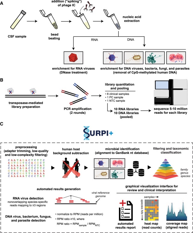
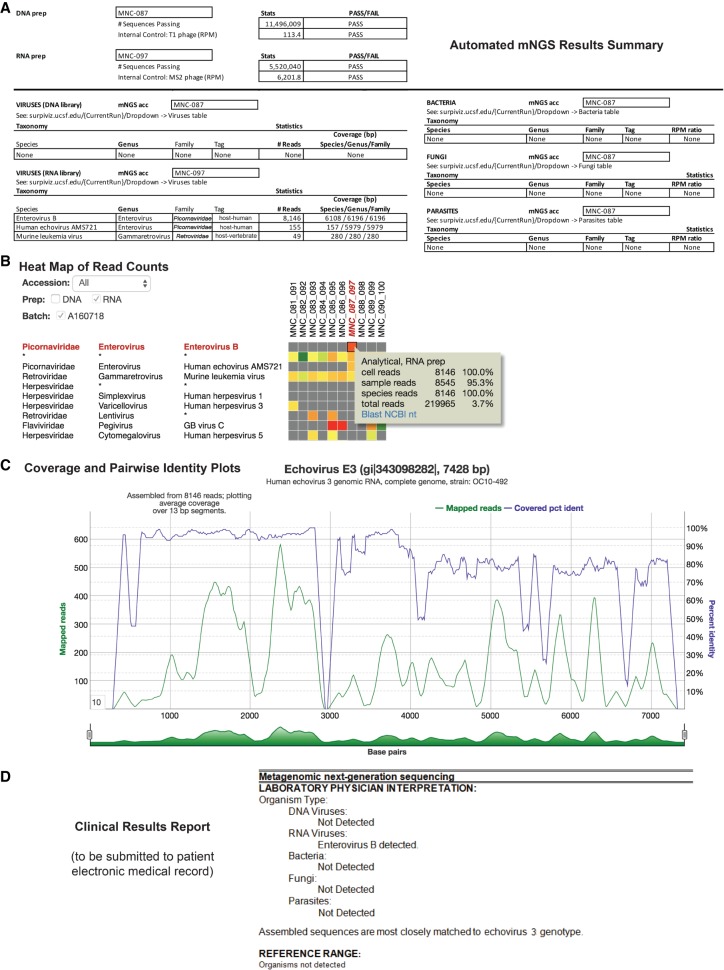
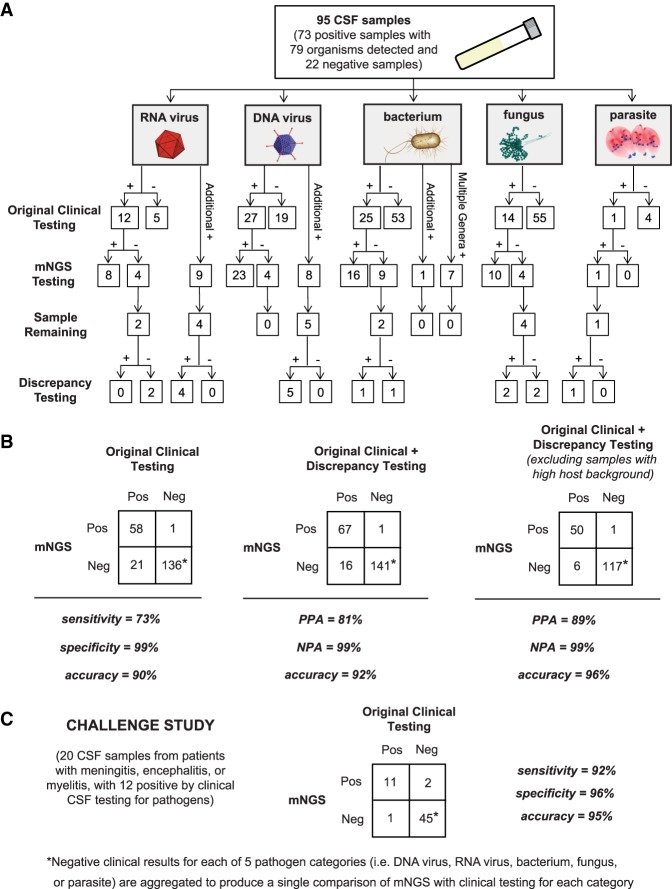
Metagenomic next-generation sequencing (mNGS) for pan-pathogen detection has been successfully tested in proof-of-concept case studies in patients with acute illness of unknown etiology but to date has been largely confined to research settings. Here, we developed and validated a clinical mNGS assay for diagnosis of infectious causes of meningitis and encephalitis from cerebrospinal fluid (CSF) in a licensed microbiology laboratory. A customized bioinformatics pipeline, SURPI+, was developed to rapidly analyze mNGS data, generate an automated summary of detected pathogens, and provide a graphical user interface for evaluating and interpreting results. We established quality metrics, threshold values, and limits of detection of 0.2-313 genomic copies or colony forming units per milliliter for each representative organism type. Gross hemolysis and excess host nucleic acid reduced assay sensitivity; however, spiked phages used as internal controls were reliable indicators of sensitivity loss. Diagnostic test accuracy was evaluated by blinded mNGS testing of 95 patient samples, revealing 73% sensitivity and 99% specificity compared to original clinical test results, and 81% positive percent agreement and 99% negative percent agreement after discrepancy analysis. Subsequent mNGS challenge testing of 20 positive CSF samples prospectively collected from a cohort of pediatric patients hospitalized with meningitis, encephalitis, and/or myelitis showed 92% sensitivity and 96% specificity relative to conventional microbiological testing of CSF in identifying the causative pathogen. These results demonstrate the analytic performance of a laboratory-validated mNGS assay for pan-pathogen detection, to be used clinically for diagnosis of neurological infections from CSF.


Figures

Figure 1.
Figure 1.
Schematic of the mNGS assay workflow. (A) CSF is extracted after lysis by bead-beating and internal control addition to allow viral, bacterial, fungal, and parasite nucleic acid retrieval. Total nucleic acid extracts are enriched for pathogen DNA by removal of methylated DNA (DNA libraries) and treatment with DNase (RNA libraries). (B) Libraries are generated using the Nextera XT protocol and amplified using two rounds of PCR. Libraries are quantified, pooled, and loaded onto the sequencer. (C) Sequences are processed using SURPI+ software for alignment and classification. Reads are preprocessed by trimming of adapters and removal of low-quality/low-complexity sequences, followed by computational subtraction of human reads and taxonomic classification of remaining microbial reads to family, genus, or species. For viruses, reads are mapped to the closest matched genome to identify nonoverlapping regions; for bacteria, fungi, and parasites, a read per million (RPM) ratio (RPM-r) metric is calculated, defined as RPM-r = RPMsample/NTC. To aid in analysis, automated result summaries, heat maps of raw/normalized read counts, and coverage/pairwise identity plots are generated for use in review and clinical interpretation.
Figure 2.
Figure 2.
Identification and reporting of enterovirus infection in a patient with meningoencephalitis using a clinical CSF mNGS assay. SURPI+ provides tools to aid clinical interpretation and graphical visualization (the SURPIviz package), including (A) an automated mNGS results summary, (B) a heat map of aligned reads corresponding to detected pathogens, and (C) a coverage plot (green line) of reads corresponding to a detected pathogen that are mapped to the most closely matched genome or gene in the reference database, along with a corresponding pairwise identity plot (purple line, sliding window = 10 nt). Viral hits corresponding to one CSF patient sample (MNC_087_097, column highlighted in red in B) are taxonomically identified as enterovirus (enterovirus B and echovirus AMS721 species) and murine leukemia virus, a known reagent contaminant (Zheng et al. 2011). After an interpretive review, a laboratory physician prepares a clinical results report (D) that is submitted to the patient electronic medical record (EMR).
Figure 3.
Figure 3.
Accuracy of mNGS relative to clinical testing of CSF. (A) Flow chart of results from samples evaluated in the accuracy study. Results are separated by organism category (RNA virus, DNA virus, bacterium, fungus, and parasite). Shown are the number of samples positive or negative by clinical testing (first row), agreement between mNGS results and positive clinical results and additional positive detections by mNGS (second row), and, in samples with sufficient remaining volume (third row), the results of orthogonal confirmatory testing (fourth row). (B) 2 × 2 contingency tables comparing the performance of mNGS relative to clinical testing of CSF. The composite reference standards used are original clinical testing (left), combined original clinical and discrepancy testing (middle), and combined original clinical and discrepancy testing after excluding high host background samples (right). (PPA) Positive predictive agreement, (NPA) negative predictive agreement. (C) 2 × 2 contingency table showing the results of challenge study. Twenty CSF samples were analyzed by mNGS and compared with the results of conventional clinical testing.

Similar articles

Cited by

References

    1. Altschul SF, Gish W, Miller W, Myers EW, Lipman DJ. 1990. Basic local alignment search tool. J Mol Biol 215: 403–410. 10.1016/S0022-2836(05)80360-2 - DOI - PubMed
    1. Bohlin J, Eldholm V, Brynildsrud O, Petterson JH, Alfsnes K. 2018. Modeling of the GC content of the substituted bases in bacterial core genomes. BMC Genomics 19: 589 10.1186/s12864-018-4984-3 - DOI - PMC - PubMed
    1. Cazanave C, Greenwood-Quaintance KE, Hanssen AD, Karau MJ, Schmidt SM, Gomez Urena EO, Mandrekar JN, Osmon DR, Lough LE, Pritt BS, et al. 2013. Rapid molecular microbiologic diagnosis of prosthetic joint infection. J Clin Microbiol 51: 2280–2287. 10.1128/JCM.00335-13 - DOI - PMC - PubMed
    1. Chiu CY. 2013. Viral pathogen discovery. Curr Opin Microbiol 16: 468–478. 10.1016/j.mib.2013.05.001 - DOI - PMC - PubMed
    1. Chiu C, Miller S. 2016. Next-generation sequencing. In Molecular microbiology: diagnostic princples and practice, 3rd ed. (ed. Persing DH, et al.), pp. 68–79. ASM Press, Washington, DC.

Publication types

MeSH terms