. 2019 May 31;294(22):8779-8790.
doi: 10.1074/jbc.RA118.007004. Epub 2019 Apr 16.

Tau repeat regions contain conserved histidine residues that modulate microtubule-binding in response to changes in pH

Affiliations

Tau repeat regions contain conserved histidine residues that modulate microtubule-binding in response to changes in pH

Rabab A Charafeddine et al. J Biol Chem. .

Abstract

Tau, a member of the MAP2/tau family of microtubule-associated proteins, stabilizes and organizes axonal microtubules in healthy neurons. In neurodegenerative tauopathies, tau dissociates from microtubules and forms neurotoxic extracellular aggregates. MAP2/tau family proteins are characterized by three to five conserved, intrinsically disordered repeat regions that mediate electrostatic interactions with the microtubule surface. Here, we used molecular dynamics, microtubule-binding experiments, and live-cell microscopy, revealing that highly-conserved histidine residues near the C terminus of each microtubule-binding repeat are pH sensors that can modulate tau-microtubule interaction strength within the physiological intracellular pH range. We observed that at low pH (<7.5), these histidines are positively charged and interact with phenylalanine residues in a hydrophobic cleft between adjacent tubulin dimers. At higher pH (>7.5), tau deprotonation decreased binding to microtubules both in vitro and in cells. Electrostatic and hydrophobic characteristics of histidine were both required for tau-microtubule binding, as substitutions with constitutively and positively charged nonaromatic lysine or uncharged alanine greatly reduced or abolished tau-microtubule binding. Consistent with these findings, tau-microtubule binding was reduced in a cancer cell model with increased intracellular pH but was rapidly restored by decreasing the pH to normal levels. These results add detailed insights into the intracellular regulation of tau activity that may be relevant in both normal and pathological conditions.

Keywords: Tau protein (Tau); cancer biology; histidine; intracellular pH; intrinsically disordered protein; microtubule; microtubule-associated protein (MAP); molecular dynamics; neurobiology; neurodegeneration; neuronal cytoskeleton; pH sensing; protein–protein interaction.


Conflict of interest statement

The authors declare that they have no conflicts of interest with the contents of this article

Figures

Figure 1.
Figure 1.
Molecular dynamics simulation of protonation effects on tau His-299 interaction with the MT surface. A, view of the MT-bound tau R2 MT-binding repeat from the MT outside based on the recent cryo-EM structure (PDB code 6CVN). The tubulin surface is colored by electrostatic potential highlighting the strongly negative surface charge or the protofilament ridge. B, last frames of 5-ns MD simulations of protonated His-299+ (i.e. low pH; orange) and unprotonated His-2990 (high pH; blue) compared with the cryo-EM structure (gray). A side view of the tubulin protofilament is shown with tau R2 on top. C, close-up views of the binding pocket surrounding His-299. D, sequence alignment of MT-binding repeats in all members of the human MAP2/tau family indicating the high degree of conservation of the histidine residue in the position equivalent to His-299 in tau R2 (asterisk).
Figure 2.
Figure 2.
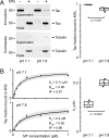
Increased pH decreases tau binding to MTs in vitro. A, co-sedimentation assay of 100 nm 0N3R tau protein with 0.5 μm paclitaxel-stabilized MTs. Shown are immunoblots for tau and Coomassie-stained gels for tubulin in the supernatant and pellet at the indicated pH values. Note that tau remains in the supernatant in the absence of MTs at both pH values. The box plot shows a quantification of the tau fraction recovered in the pellet (n = 5). B, equilibrium binding of 50 nm fluorescently tagged 0N3R sfGFP–tau protein at a range of MT concentrations between 15.6 nm and 1 μm at the indicated pH values. The fraction bound was determined by MT co-sedimentation of fluorescently-tagged 0N3R sfGFP–tau and measuring sfGFP–tau fluorescence in the supernatant and pellet. Gray symbols show each data point, and black symbols are the average for each MT concentration. The black line shows a hyperbolic fit through data from all experiments, and the gray area indicates the 95% confidence interval of the fit. The box plot shows dissociation constants obtained when each experiment was fitted independently (n = 4). Box plots show median, first, and third quartile, with whiskers extending to observations within 1.5 times the interquartile range, and all individual data points. Statistical analysis was by Student's t test.
Figure 3.
Figure 3.
Increased intracellular pH decreases tau binding to MTs. A, RPE cells transiently expressing either fluorescently tagged 0N3R tau (top row) or tubulin (bottom row) treated with 20 mm NH4Cl to acutely increase pHi in the cytoplasm. Insets show highlighted regions at higher magnification. B, quantification of the mEmerald–tau or EGFP-tubulin fluorescence in the cytoplasm (n = 13 cells each). Note that elevated pHi increases mEmerald–tau signal in the cytoplasm almost 2-fold, which indicates dissociation from MTs. Box plots show median, first, and third quartile, with whiskers extending to observations within 1.5 times the interquartile range, and all individual data points. Statistical analysis was by Tukey-Kramer HSD test.
Figure 4.
Figure 4.
Increased intracellular pH decreases binding of MAP2/tau family proteins to MTs. A, RPE cells transiently expressing either mEmerald-tagged tau (top row) or -MAP4 (bottom row) treated with 100 mm NaCl to acutely increase pHi in the cytoplasm. Insets show highlighted regions at higher magnification. B, quantification of the mEmerald–tau or -MAP4 fluorescence in the cytoplasm (tau, n = 19; MAP4, n = 13 cells). Both MAP2/tau family proteins reversibly dissociate from MTs at increased pHi. Note that MAP4 expression generally results in some degree of MT bundling, and MAP4 does not completely disappear from these bundles, but mEmerald-MAP4 in the cytoplasm is notably increased at increased pHi values. Box plots show median, first, and third quartile, with whiskers extending to observations within 1.5 times the interquartile range, and all individual data points. Statistical analysis was by Tukey-Kramer HSD test.
Figure 5.
Figure 5.
Decreasing pHi in cancer cells enhances tau binding to MTs. A, MCF10A mammary epithelial cells expressing oncogenic H-RasV12 before and after treatment with a nigericin buffer to equilibrate pHi values to 7.2. Inset shows highlighted regions at higher magnification. B, quantification of the relative mEmerald–tau fluorescence in the cytoplasm (n = 15 cells). Box plot shows median, first, and third quartile, whiskers extending to observations within 1.5 times the interquartile range, and all individual data points. Statistical analysis was by Student's t test.
Figure 6.
Figure 6.
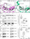
Conserved histidine residues modulate pH-dependent tau binding to MTs. A, close-up views of 5-ns MD simulations in which tau R2 His-299 was substituted with either lysine or alanine as indicated overlaid with the cryo-EM structure shown in gray. B, co-sedimentation assay of 100 nm 0N3R tau protein with the indicated substitutions of conserved histidine residues with 1 μm paclitaxel-stabilized MTs at pH 6.8. Shown are immunoblots for tau and Coomassie-stained gels for tubulin in the supernatant and pellet. Note that tau remains in the supernatant in the absence of MTs. The box plot shows a quantification of the tau fraction recovered in the pellet (n = 5). C, Co-sedimentation assay of 10 nm 0N3R tau protein or 10 nm tau(H4K) with 0.5 μm paclitaxel-stabilized MTs. Because tau is too diluted to be accurately detected in the supernatant, only pellets of two independent experiments for each tau construct at the indicated pH values are shown. Equivalent amounts of pellets from different experiments were run in adjacent lanes on the same gels to minimize experimental error. The box plot shows the ratio of either tubulin or tau in the pellet at pH 7.8 compared with pH 7.1 (WT, n = 4; H4K, n = 3). Statistical analysis was by Tukey-Kramer HSD test.
Figure 7.
Figure 7.
Conserved histidine residues are required for tau binding to MTs in cells. A, RPE cells transiently expressing the indicated mEmerald-tagged tau constructs. Insets show highlighted regions at higher magnification. B, quantification of the relative enrichment of the indicated mEmerald–tau constructs on MTs compared with cytoplasm signal (WT and H4K: n = 14; H4A: n = 6 cells). The dashed line at a MT to cytoplasm ratio of 1 represents undetectable MT binding. Box plot shows median, first, and third quartile, with whiskers extending to observations within 1.5 times the interquartile range, and all individual data points. Statistical analysis was by Tukey-Kramer HSD test.

Similar articles

Cited by

References

    1. Dehmelt L., and Halpain S. (2005) The MAP2/Tau family of microtubule-associated proteins. Genome Biol. 6, 204 10.1186/gb-2004-6-1-204 - DOI - PMC - PubMed
    1. Al-Bassam J., Ozer R. S., Safer D., Halpain S., and Milligan R. A. (2002) MAP2 and tau bind longitudinally along the outer ridges of microtubule protofilaments. J. Cell Biol. 157, 1187–1196 10.1083/jcb.200201048 - DOI - PMC - PubMed
    1. Wang Y., and Mandelkow E. (2016) Tau in physiology and pathology. Nat. Rev. Neurosci. 17, 5–21 10.1038/nrg.2015.11 - DOI - PubMed
    1. Qiang L., Yu W., Andreadis A., Luo M., and Baas P. W. (2006) Tau protects microtubules in the axon from severing by katanin. J. Neurosci. 26, 3120–3129 10.1523/JNEUROSCI.5392-05.2006 - DOI - PMC - PubMed
    1. Dixit R., Ross J. L., Goldman Y. E., and Holzbaur E. L. (2008) Differential regulation of dynein and kinesin motor proteins by tau. Science 319, 1086–1089 10.1126/science.1152993 - DOI - PMC - PubMed

Publication types

Associated data