. 2019 Apr 16;12(577):eaau9685.
doi: 10.1126/scisignal.aau9685.

Cardiomyocyte glucocorticoid and mineralocorticoid receptors directly and antagonistically regulate heart disease in mice

Affiliations

Cardiomyocyte glucocorticoid and mineralocorticoid receptors directly and antagonistically regulate heart disease in mice

Robert H Oakley et al. Sci Signal. .

Abstract

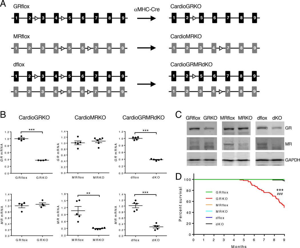
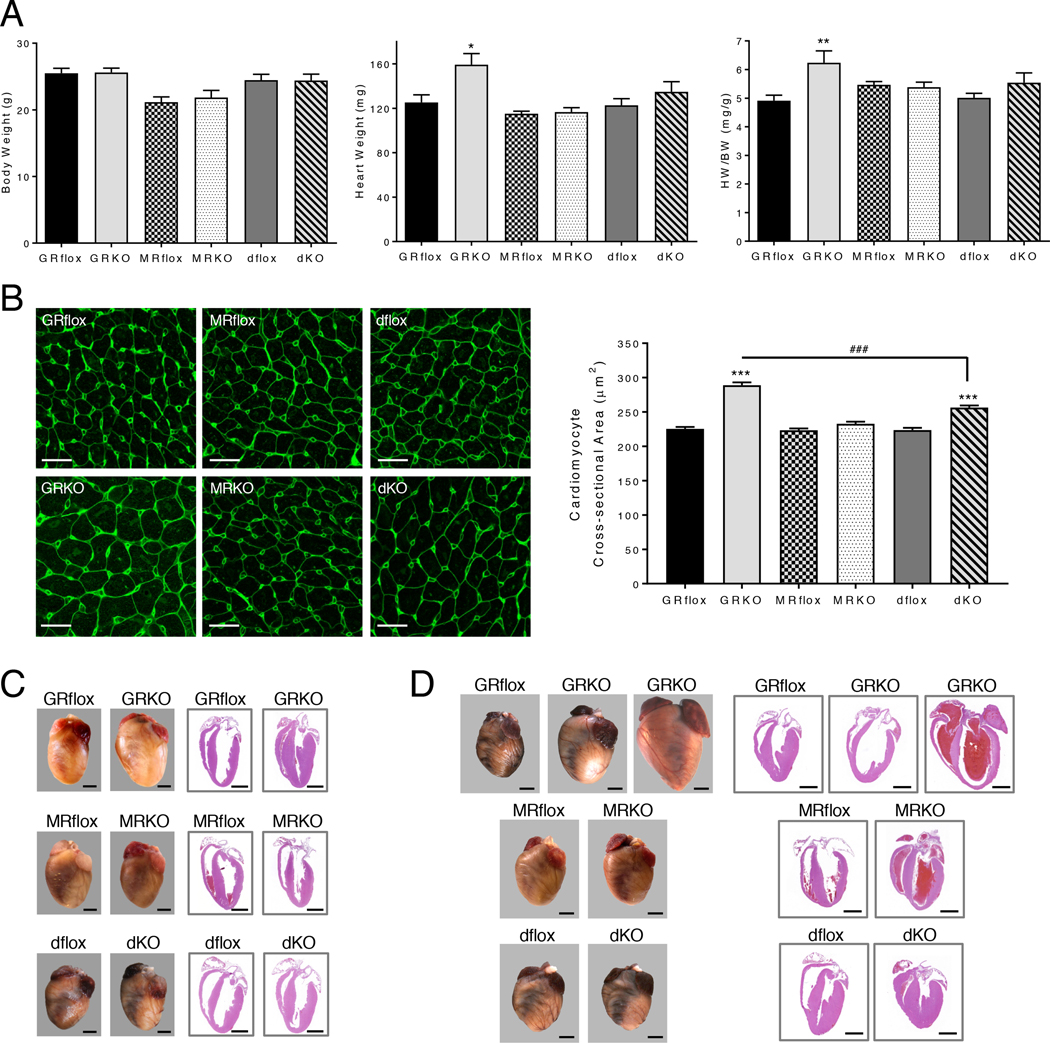
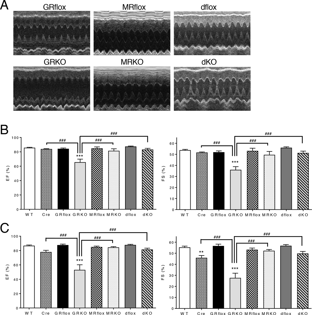
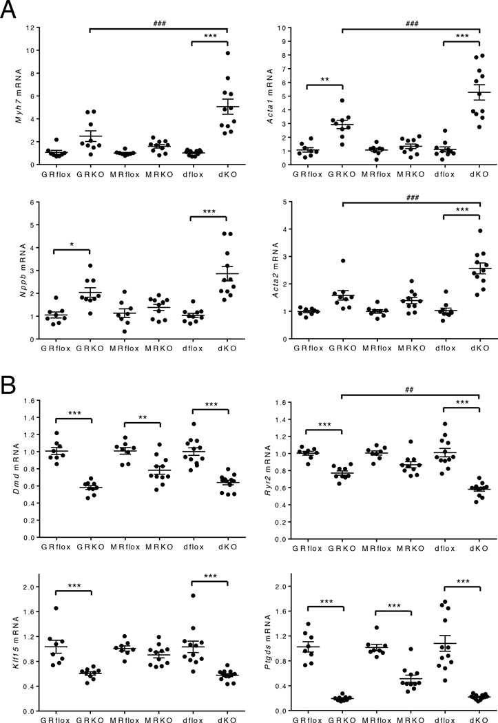
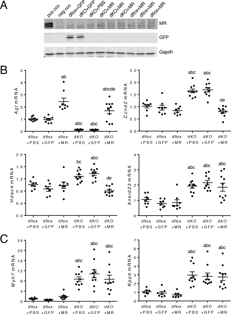
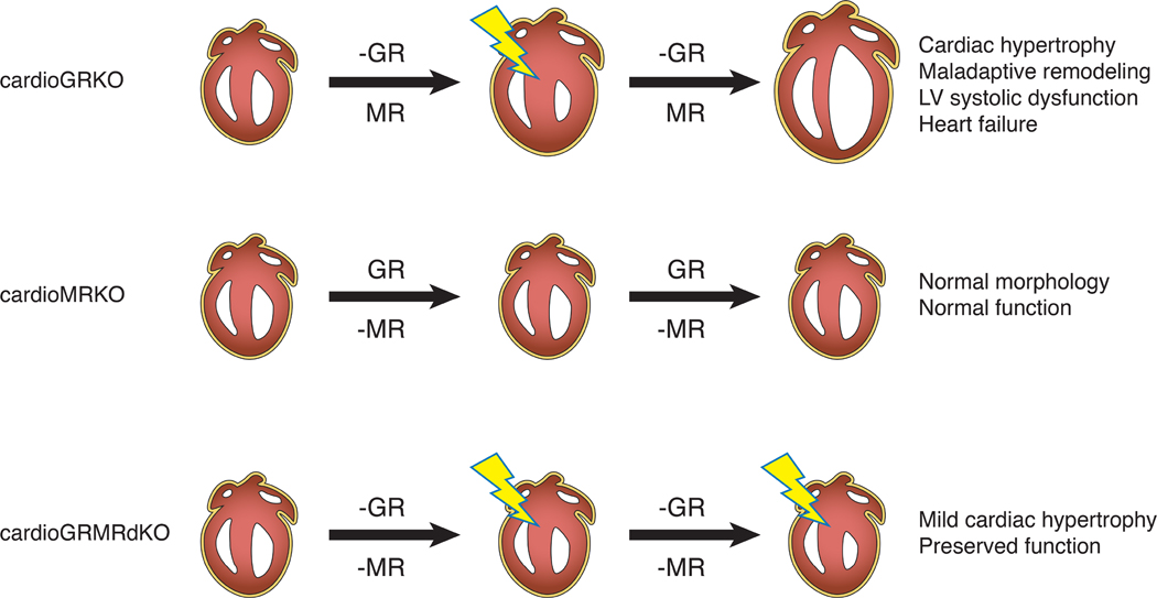
Stress is increasingly associated with heart dysfunction and is linked to higher mortality rates in patients with cardiometabolic disease. Glucocorticoids are primary stress hormones that regulate homeostasis through two nuclear receptors, the glucocorticoid receptor (GR) and mineralocorticoid receptor (MR), both of which are present in cardiomyocytes. To examine the specific and coordinated roles that these receptors play in mediating the direct effects of stress on the heart, we generated mice with cardiomyocyte-specific deletion of GR (cardioGRKO), MR (cardioMRKO), or both GR and MR (cardioGRMRdKO). The cardioGRKO mice spontaneously developed cardiac hypertrophy and left ventricular systolic dysfunction and died prematurely from heart failure. In contrast, the cardioMRKO mice exhibited normal heart morphology and function. Despite the presence of myocardial stress, the cardioGRMRdKO mice were resistant to the cardiac remodeling, left ventricular dysfunction, and early death observed in the cardioGRKO mice. Gene expression analysis revealed the loss of gene changes associated with impaired Ca2+ handling, increased oxidative stress, and enhanced cell death and the presence of gene changes that limited the hypertrophic response and promoted cardiomyocyte survival in the double knockout hearts. Reexpression of MR in cardioGRMRdKO hearts reversed many of the cardioprotective gene changes and resulted in cardiac failure. These findings reveal a critical role for balanced cardiomyocyte GR and MR stress signaling in cardiovascular health. Therapies that shift stress signaling in the heart to favor more GR and less MR activity may provide an improved approach for treating heart disease.


Conflict of interest statement

Competing interests: The authors have declared that no conflict of interest exists.

Figures

Figure 1.
Figure 1.. Generation and survival of mice with conditional knockout of GR, MR, or both GR and MR in cardiomyocytes.
(A) Mice deficient in cardiomyocyte GR (cardioGRKO), MR (cardioMRKO), or both GR and MR (cardioGRMRdKO) were generated by crossing mice with floxed GR and/or MR alleles with mice expressing Cre recombinase only in cardiomyocytes (αMHC-Cre). (B) RT-PCR of GR and MR mRNA from hearts of 2-month old knockout mice and their littermate controls. Data are mean ± SEM (n = 4–6 mice per group). Student’s t test was performed to determine significance. **P < 0.01 and ***P < 0.001 for GRKO compared to GRflox, for MRKO compared to MRflox, and for dKO compared to dflox. (C) Representative immunoblots of GR and MR protein from hearts of 3-month old knockout mice and their littermate controls (n = 3 independent experiments). (D) Survival curves for GRflox (n = 36), cardioGRKO (n = 92), MRflox (n = 26), cardioMRKO (n = 54), dflox (n = 41), and cardioGRMRdKO (n = 85) mice. Mantel-Cox log-rank test was performed with a Bonferroni corrected threshold to determine significance. ***P < 0.001 for GRKO compared to GRflox. ###P < 0.001 for MRKO compared to GRKO and for dKO compared to GRKO.
Figure 2.
Figure 2.. CardioGRMRdKO mice are protected from LV remodeling.
(A) Body weight (BW), heart weight (HW), and HW/BW ratios were determined for 2-month old control and knockout mice. Data are mean ± SEM (n = 5–9 mice per group). (B) Cross-sectional area of LV cardiomyocytes in 2-month old control and knockout hearts. Left panel shows representative confocal images of heart sections stained with FITC-lectin. Scale bar is 20 μm. Right panel shows quantitation of cardiomyocyte cross-sectional area. Data are mean ± SEM (greater than 400 cardiomyocytes from n = 4–5 mice per group). (C, D) Representative images of intact hearts (left panel) and longitudinal H&E-stained heart sections (right panel) from 3-month (C) and 6-month (D) old control and knockout mice. Scale bar is 2mm. Images are representative of 3–6 mice per genotype. A one-way ANOVA was performed to determine significance. *P < 0.05, **P < 0.01, and ***P < 0.001 for GRKO compared to GRflox and for dKO compared to dflox. ###P < 0.001 for dKO compared to GRKO.
Figure 3.
Figure 3.. CardioGRMRdKO mice are protected from LV systolic dysfunction.
(A) Representative M-mode images from 3-month old control and knockout mice. (B, C) Echocardiographic measurements of percent ejection fraction (EF) and percent fractional shortening (FS) were determined from transthoracic M-mode tracings made on control and knockout mice that were 3 months (B) or 6 months (C) old. Data are mean ± SEM (n = 5–11 mice per group). A one-way ANOVA was performed to determine significance. **P < 0.01 and ***P < 0.001 for GRKO compared to GRflox and for Cre compared to WT. ###P < 0.001 for Cre compared to GRKO, for MRKO compared to GRKO, and for dKO compared to GRKO.
Figure 4.
Figure 4.. Genes associated with cardiac pathology are dysregulated in cardioGRMRdKO hearts.
Total RNA was isolated from whole hearts from 2-month old control and knockout mice. (A) Myh7, Acta1, Nppb, and Acta2 mRNA levels were measured by RTPCR. Data are mean ± SEM (n = 8–11 mice per group). (B) Dmd, Ryr2, Klf15, and Ptgds mRNA levels were measured by RTPCR. Data are mean ± SEM (n = 8–12 mice per group). A one-way ANOVA was performed to determine significance. *P < 0.05, **P < 0.01, and ***P < 0.001 for GRKO compared to GRflox, for MRKO compared to MRflox, and for dKO compared to dflox. ##P < 0.01 and ###P < 0.001 for dKO compared to GRKO.
Figure 5.
Figure 5.. Global gene expression profile in 1-month old cardioGRKO, cardioMRKO, and cardioGRMRdKO hearts.
Microarrays were performed on RNA isolated from the hearts of 1-month old control and knockout mice. (A) Total number of genes differentially expressed in the hearts of knockout mice compared to their control littermates. (B) Differentially expressed genes in the knockout hearts were compared using a Venn diagram. (C) Diseases and disorders most significantly associated with the dysregulated genes in the knockout hearts as determined by IPA. (D) Gene enrichment comparison analysis of the dysregulated genes associated with “Cardiovascular Disease” in the 3 knockout hearts was performed using IPA. Shown are the disease annotations with a significant activation z-score (absolute value ≥ 2). The retrieved annotation “Failure of Heart” was only significantly associated with the dysregulated genes in the cardioGRKO heart. ns = not significant.
Figure 6.
Figure 6.. CardioGRMRdKO hearts are protected from alterations in Ca2+ handling and oxidative stress observed in cardioGRKO hearts.
(A) Signaling pathways most significantly associated with the dysregulated genes in 1-month old knockout hearts as determined by IPA. (B) The “Cardiac β-Adrenergic Signaling” pathway overlaid with dysregulated genes in 1-month old knockout hearts. Red and green colors correspond to up-regulation and down-regulation, respectively. (C, D) mRNA levels for the Ca2+ handling genes Cacna1c, Atp2a2, and Slc8a1 (C) and the oxidative stress genes Ncf1, Rac2, and Spp1 (D) were measured by RTPCR in 3-month old control and knockout hearts. Data are mean ± SEM (n = 7–10 mice per group). *P < 0.05 and ***P < 0.001 for GRKO compared to GRflox, for MRKO compared to MRflox, and for dKO compared to dflox.
Figure 7.
Figure 7.. CardioGRMRdKO hearts are protected from cell death observed in cardioGRKO hearts.
(A) Molecular and cellular functions most significantly associated with the dysregulated genes in 1-month old knockout hearts as determined by IPA. (B) Dysregulated genes associated with “Cell Death and Survival” in the knockout hearts were compared using a Venn diagram. (C) Gene enrichment comparison analysis of the dysregulated genes associated with “Cell Death and Survival” in the 3 knockout hearts was performed using IPA. Shown are the functional annotations with a significant activation z-score (absolute value ≥ 2). The retrieved annotations were only significantly associated with the dysregulated genes in the cardioGRKO heart. ns = not significant. (D) Analysis of cell death in knockout hearts. Left panel shows representative image of TUNEL-positive nuclei (arrow) in LV myocardium of 6-month cardioGRKO heart. Quadruple staining was performed: TUNEL (green), WGA (red), cardiac troponin T (Tnnt2) (cyan), and DAPI (blue). Scale bar is 10 μm. Right panel shows quantitation of TUNEL-positive nuclei in 6-month knockout hearts. Data are mean ± SEM (n = 4–6 mice per group). A one-way ANOVA was performed to determine significance. *P < 0.05 for GRKO compared to GRflox.
Figure 8.
Figure 8.. Gene changes associated with cardioprotection are uniquely observed in cardioGRMRdKO hearts.
Total RNA was isolated from whole hearts of 1-month (A) and 2-month (B) old control and knockout hearts. Agt, Ccnd2, Hdac4, and Ankrd23 mRNA levels were measured by RTPCR. Data are mean ± SEM (n = 4–9 mice per group). A one-way ANOVA was performed to determine significance. *P < 0.05, **P < 0.01, and ***P < 0.001 for GRKO compared to GRflox, for MRKO compared to MRflox, and for dKO compared to dflox. #P < 0.05, ##P < 0.01, and ###P < 0.001 for dKO compared to GRKO. &P < 0.05, &&P < 0.01, and &&&P < 0.001 for dKO compared to MRKO.
Figure 9.
Figure 9.. Re-expression of MR in the cardioGRMRdKO heart reverses cardioprotective gene changes.
CardioGRMRdKO mice and their littermate controls (dflox) were injected intravenously with PBS, AAV-Tnnt2-GFP, or AAV-Tnnt2-MR at 4–6 weeks of age. (A) Representative immunoblot shows MR and GFP expression in hearts isolated from injected mice that were 6 months old (n = 3 independent experiments). Positive (pos) and negative (neg) controls are hippocampal lysates from a wild-type mouse and a littermate mouse with conditional knockout of MR in the hippocampus, respectively. (B) RTPCR analysis of Agt, Ccnd2, Hdac4, and Ankrd23 mRNA levels in hearts from injected dflox and cardioGRMRdKO mice that were 6 months old. Data are mean ± SEM (n = 7–10 mice per group). (C) RTPCR analysis of Myh7 and Nppb mRNA levels in hearts from injected dflox and cardioGRMRdKO mice that were 6 months old. Data are mean ± SEM (n = 7–10 mice per group). A one-way ANOVA was performed to determine significance. aP < 0.05 compared to dflox+PBS. bP < 0.05 compared to dflox+GFP. cP < 0.05 compared to dflox+MR. dP < 0.05 compared to dKO+PBS. eP < 0.05 compared to dKO+GFP.
Figure 10.
Figure 10.. Cardiomyocyte GR and MR signaling and heart disease.
Findings from our genetic mouse models suggest that both insufficient cardiomyocyte GR signaling and deleterious cardiomyocyte MR signaling contribute to heart disease. A deficiency in cardiomyocyte GR signaling alone (-GR) leads to myocardial stress (lightning bolt) and mild hypertrophy in both the cardioGRKO and cardioGRMRdKO hearts. In contrast, a deficiency in cardiomyocyte MR signaling alone (-MR) does not have an overt effect. In the cardioGRKO hearts, cardiomyocyte MR signaling becomes deleterious and exacerbates the hypertrophic response leading to maladaptive remodeling and heart failure. In the cardioGRMRdKO hearts, the mild cardiac hypertrophy triggered by the loss of cardiomyocyte GR signaling is not exacerbated in the absence of cardiomyocyte MR signaling and heart function is preserved for a longer period of time.

Comment in

  • Balancing stress signalling in the heart.
    Fernández-Ruiz I. Fernández-Ruiz I. Nat Rev Cardiol. 2019 Jul;16(7):384-385. doi: 10.1038/s41569-019-0207-0. Nat Rev Cardiol. 2019. PMID: 31068688 No abstract available.

Similar articles

Cited by

References

    1. Brotman DJ, Golden SH, Wittstein IS, The cardiovascular toll of stress. Lancet 370, 1089–1100 (2007). - PubMed
    1. Steptoe A, Kivimaki M, Stress and cardiovascular disease. Nature reviews. Cardiology 9, 360–370 (2012). - PubMed
    1. Kivimaki M, Pentti J, Ferrie JE, Batty GD, Nyberg ST, Jokela M, Virtanen M, Alfredsson L, Dragano N, Fransson EI, Goldberg M, Knutsson A, Koskenvuo M, Koskinen A, Kouvonen A, Luukkonen R, Oksanen T, Rugulies R, Siegrist J, Singh-Manoux A, Suominen S, Theorell T, Vaananen A, Vahtera J, Westerholm PJM, Westerlund H, Zins M, Strandberg T, Steptoe A, Deanfield J, consortium IP-W, Work stress and risk of death in men and women with and without cardiometabolic disease: a multicohort study. Lancet Diabetes Endocrinol, (2018). - PMC - PubMed
    1. Stewart RAH, Colquhoun DM, Marschner SL, Kirby AC, Simes J, Nestel PJ, Glozier N, O’Neil A, Oldenburg B, White HD, Tonkin AM, Investigators LS, Persistent psychological distress and mortality in patients with stable coronary artery disease. Heart 103, 1860–1866 (2017). - PubMed
    1. Oakley RH, Cidlowski JA, Glucocorticoid signaling in the heart: A cardiomyocyte perspective. J Steroid Biochem Mol Biol 153, 27–34 (2015). - PMC - PubMed

Publication types

MeSH terms