. 2019 Apr 16;9(1):6171.
doi: 10.1038/s41598-019-42367-8.

RNA recognition motifs of disease-linked RNA-binding proteins contribute to amyloid formation

Affiliations

RNA recognition motifs of disease-linked RNA-binding proteins contribute to amyloid formation

Sashank Agrawal et al. Sci Rep. .

Abstract

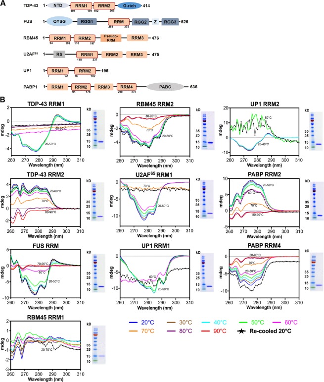
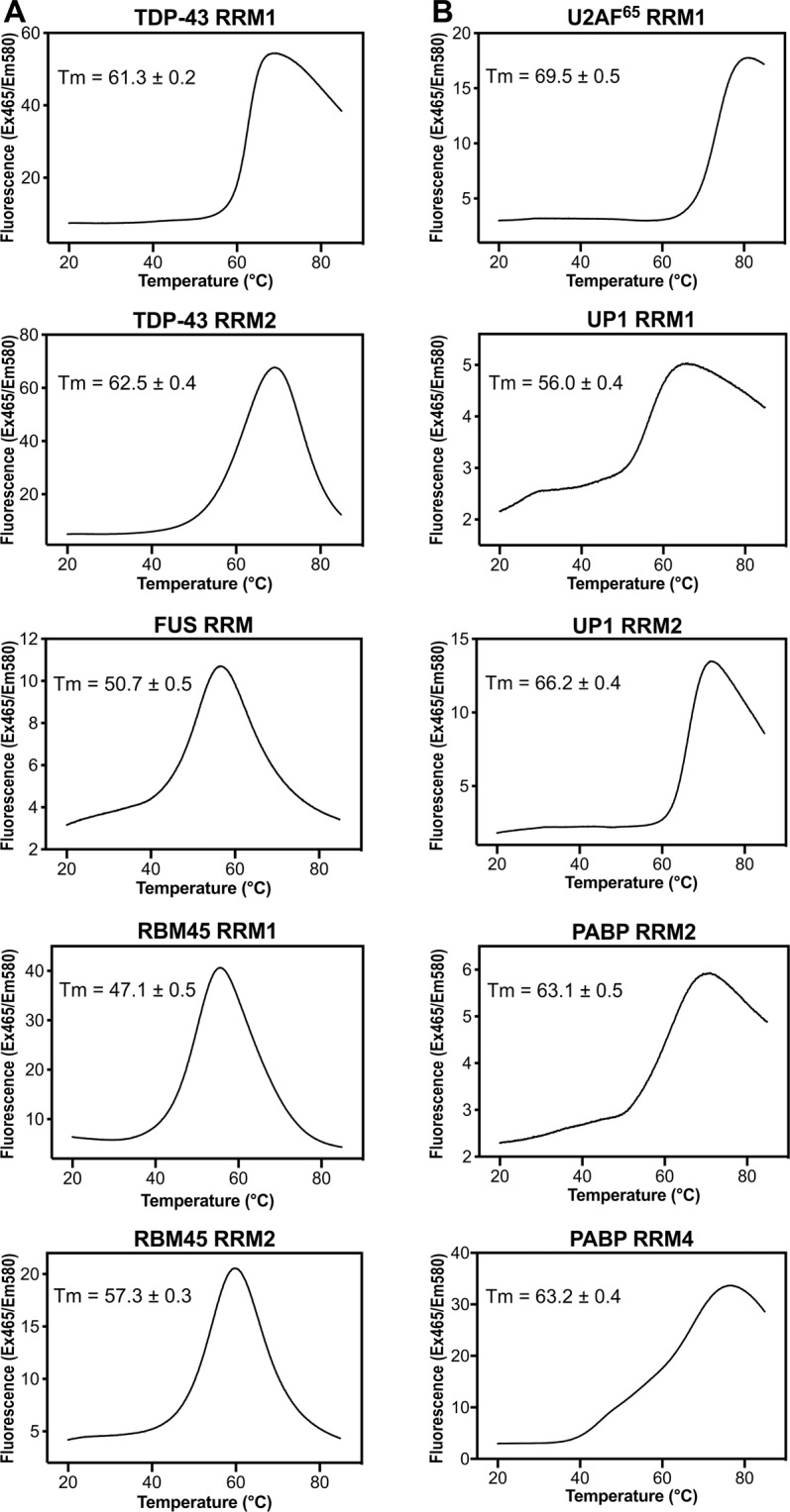
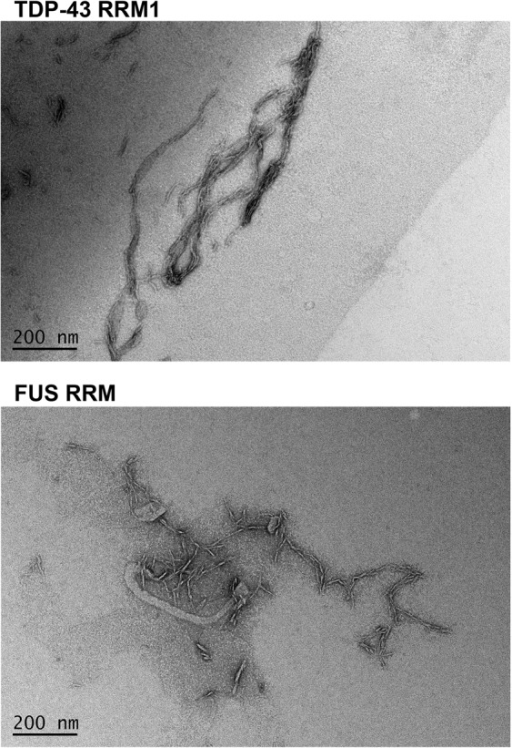
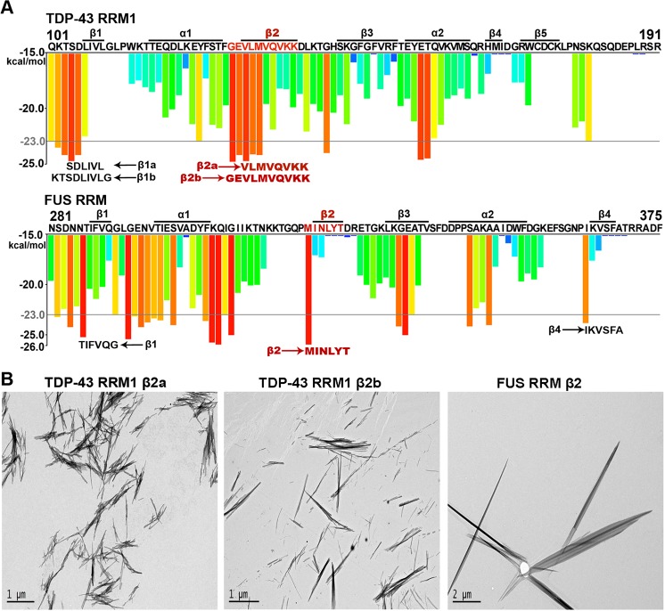
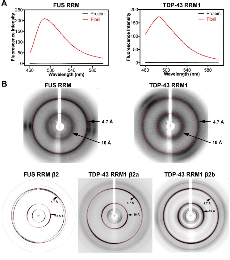
Aberrant expression, dysfunction and particularly aggregation of a group of RNA-binding proteins, including TDP-43, FUS and RBM45, are associated with neurological disorders. These three disease-linked RNA-binding proteins all contain at least one RNA recognition motif (RRM). However, it is not clear if these RRMs contribute to their aggregation-prone character. Here, we compare the biophysical and fibril formation properties of five RRMs from disease-linked RNA-binding proteins and five RRMs from non-disease-associated proteins to determine if disease-linked RRMs share specific features making them prone to self-assembly. We found that most of the disease-linked RRMs exhibit reversible thermal unfolding and refolding, and have a slightly lower average thermal melting point compared to that of normal RRMs. The full domain of TDP-43 RRM1 and FUS RRM, as well as the β-peptides from these two RRMs, could self-assemble into fibril-like aggregates which are amyloids of parallel β-sheets as verified by X-ray diffraction and FT-IR spectroscopy. Our results suggest that some disease-linked RRMs indeed play important roles in amyloid formation and shed light on why RNA-binding proteins with RRMs are frequently identified in the cellular inclusions of neurodegenerative diseases.


Conflict of interest statement

The authors declare no competing interests.

Figures

Figure 1
Figure 1
Most disease-linked RNA recognition motifs (RRMs) exhibit reversible thermal unfolding and refolding. (A) Domain organization of six RNA-binding proteins shows the ten RRMs (outlined in red box with residue numbers marked at the bottom) used in this study. (B) Overall tertiary structural changes for five RRMs from disease-linked RNA-binding proteins (TDP-43 RRM1, TDP-43 RRM2, FUS RRM, RBM45 RRM1, RBM45 RRM2), and five RRMs from non-disease-associated proteins (U2AF RRM1, UP1 RRM1, UP1 RRM2, PABP RRM2, PABP RRM4), assessed by circular dichroism in the near-UV range (260 to 310 nm) during the thermal unfolding and refolding process. We raised the temperature from 20 °C to 90 °C at intervals of 10 °C (marked by different colors, as shown at the bottom of the figure) to induce protein unfolding, and then re-cooled to 20 °C for protein refolding (marked by lines of black asterisks, labeled as “Re-cooled 20 °C”). Each RRM was purified to a high homogeneity, as shown by the SDS-PAGE gels at right of the CD spectra (The full-length gels are shown in the Supplementary Fig. S1).
Figure 2
Figure 2
Thermal melting points of disease-linked and non-disease-associated RRMs, measured by differential scanning fluorimetry (DSF). Thermal melting points of RRMs were analyzed by DSF using SYPRO orange dye. The temperature was increased from 20 °C to 85 °C at a rate of 0.06 °C/second, and the emitted SYPRO orange fluorescence signals (excited at 465 nm) were recorded at 580 nm. The thermal melting points (Tm) for (A) the disease-linked RRMs (TDP-43 RRM1, TDP-43 RRM2, FUS RRM, RBM45 RRM1, RBM45 RRM2), and for (B) non-disease-associated RRMs (U2AF RRM1, UP1 RRM1, UP1 RRM2, PABP RRM2, PABP RRM4) are shown in each panel.
Figure 3
Figure 3
RRMs from TDP-43 and FUS form fibrillar aggregates. Solutions of TDP-43 RRM1 (50 μM) and FUS RRM (50 μM) were agitated in the presence of 10 mM phosphate (pH 7.5) and 50 mM NaCl in room temperature. The freshly-formed fibrillar solutions were examined under negative-stain transmission electron microscopy (EM) to reveal fibril-like aggregates.
Figure 4
Figure 4
The β2 peptides of TDP-43 RRM1 and FUS RRM are prone to fibril formation. (A) Amyloid-promoting segments (predicted by ZipperDB) in TDP-43 RRM1 and FUS RRM are presented as red and orange bars. Amino acid sequences are listed at the top, with secondary structures (labeled as α and β) derived from the crystal structure of TDP-43 RRM1 (PDB entry: 4Y0F) and the NMR structure of FUS RRM (PDB entry: 2LCW) shown above. Three β2 peptides that formed fibrils are labeled in red, whereas the peptides that did not form fibrils are labeled in black, shown at the bottom of each histogram. (B) The peptide sequences from the β2 region of TDP-43 RRM1 (β2a and β2b) and FUS RRM (β2) form fibrils in vitro, as revealed by negative-stain transmission electron microscopy.
Figure 5
Figure 5
Fibril aggregates of TDP-43 RRM1, FUS RRM and their β2 peptides exhibit amyloid properties. (A) Fluorescence spectra (460 to 600 nm, red lines) of the freshly-formed fibrillar solutions of FUS RRM and TDP-43 RRM1 revealed fluorescence signals in the presence of Thioflavin T (excited at 442 nm), but the fresh protein solutions did not generate any signal (black lines). (B) X-ray diffraction images of the fibrils formed by FUS RRM, TDP-43 RRM1 and their β2 peptides (TDP-43 RRM1 β2a, TDP-43 RRM1 β2b and FUS RRM β2) reveal the characteristic cross-β diffraction patterns of amyloid fibrils at 4.7 Å and 10 Å (marked by red rings).
Figure 6
Figure 6
RRM and β peptide fibrils are amyloids of parallel β-sheets based on Fourier transform infrared (FTIR) spectroscopy. The fibrillation properties of FUS RRM, TDP-43 RRM1 and their β2 peptides were analyzed by attenuated total reflection-Fourier transform infrared (ATR-FTIR). ATR-FTIR spectra in the amide I region (1600 to 1700 cm−1) are shown in the left column, and the second derivative ATR-FTIR spectra are displayed in the right column. In these ATR-FTIR spectra, the characteristic shift in β-sheet absorbance at 1630–1640 cm−1 for fresh protein/peptide (in black) to 1620–1630 cm−1 in amyloid fibrils (in red) represents planer extended β-sheet assembly. In the second derivative ATR-FTIR spectra, none of the fibril spectra exhibit a high frequency peak at 1685–1695 cm−1, suggesting the presence of amyloid structures consisting of parallel β-sheets.

References

    1. Ugras SE, Shorter J. RNA-binding proteins in amyotrophic lateral sclerosis and neurodegeneration. Neurol Res Int. 2012;2012:432780. doi: 10.1155/2012/432780. - DOI - PMC - PubMed
    1. Lukong KE, Chang K-w, Khandjian EW, Richard Sp. RNA-binding proteins in human genetic disease. Trends Genet. 2008;24:416–425. doi: 10.1016/j.tig.2008.05.004. - DOI - PubMed
    1. Neumann M, et al. Ubiquitinated TDP-43 in frontotemporal lobar degeneration and amyotrophic lateral sclerosis. Science. 2006;314:130–3. doi: 10.1126/science.1134108. - DOI - PubMed
    1. Arai T, et al. TDP-43 is a component of ubiquitin-positive tau-negative inclusions in frontotemporal lobar degeneration and amyotrophic lateral sclerosis. Biochem Biophys Res Commun. 2006;351:602–11. doi: 10.1016/j.bbrc.2006.10.093. - DOI - PubMed
    1. Vance C, et al. Mutations in FUS, an RNA processing protein, cause familial amyotrophic lateral sclerosis type 6. Science. 2009;323:1208–1211. doi: 10.1126/science.1165942. - DOI - PMC - PubMed