Review
. 2019 Jul;20(7):406-420.
doi: 10.1038/s41580-019-0126-2.

Quality and quantity control of gene expression by nonsense-mediated mRNA decay

Affiliations
Review

Quality and quantity control of gene expression by nonsense-mediated mRNA decay

Tatsuaki Kurosaki et al. Nat Rev Mol Cell Biol. 2019 Jul.

Erratum in

Abstract

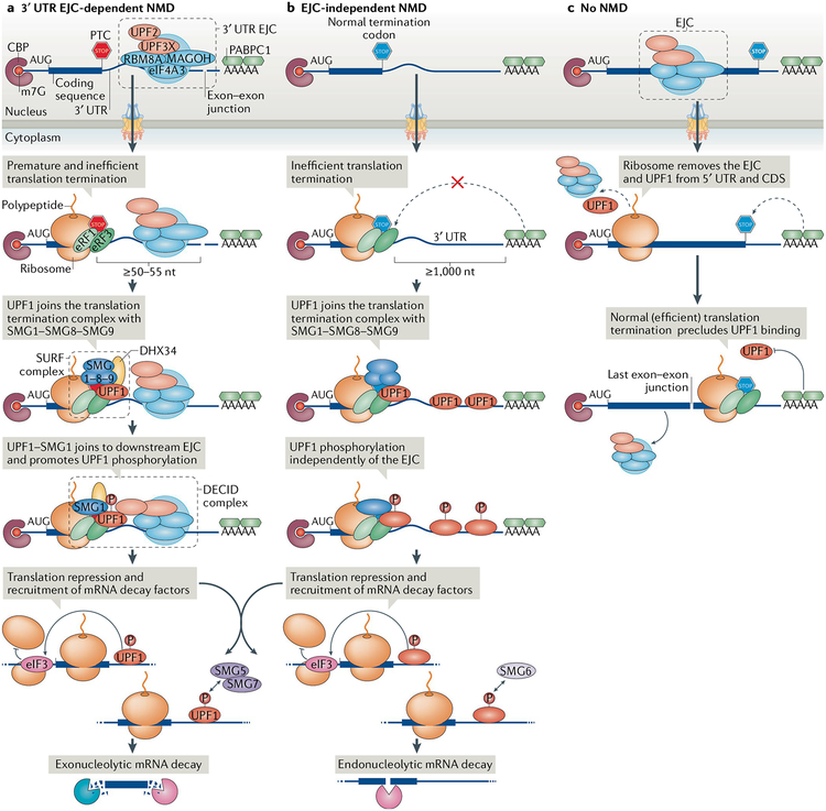
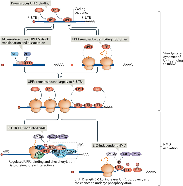
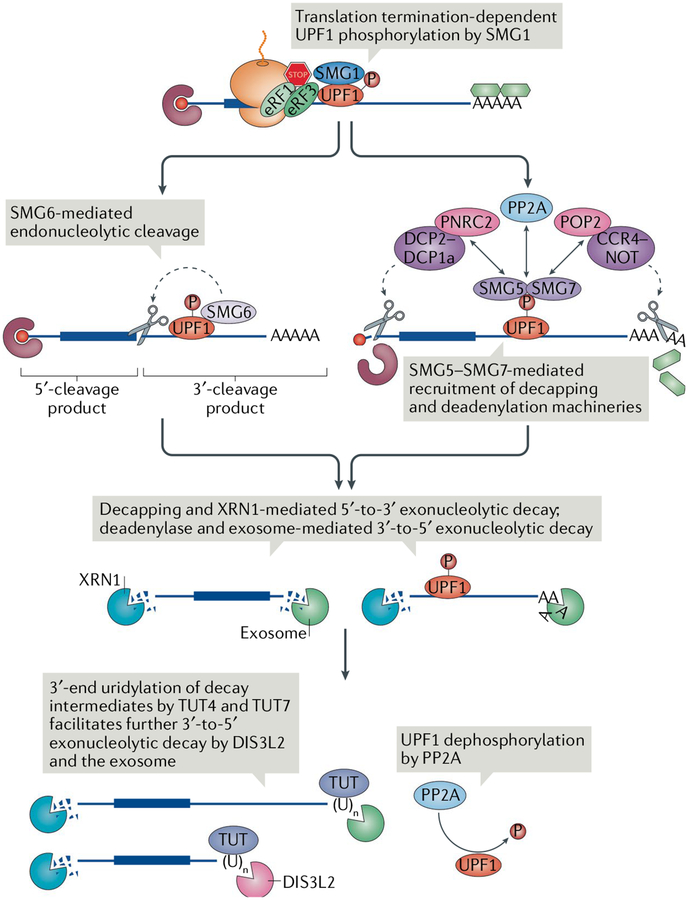
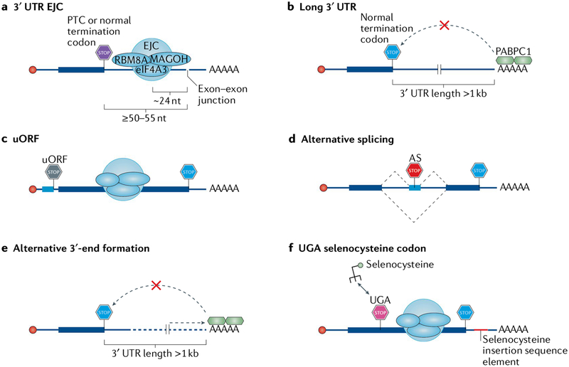
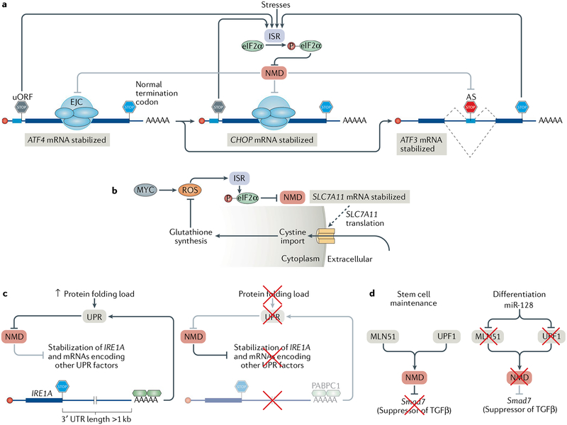
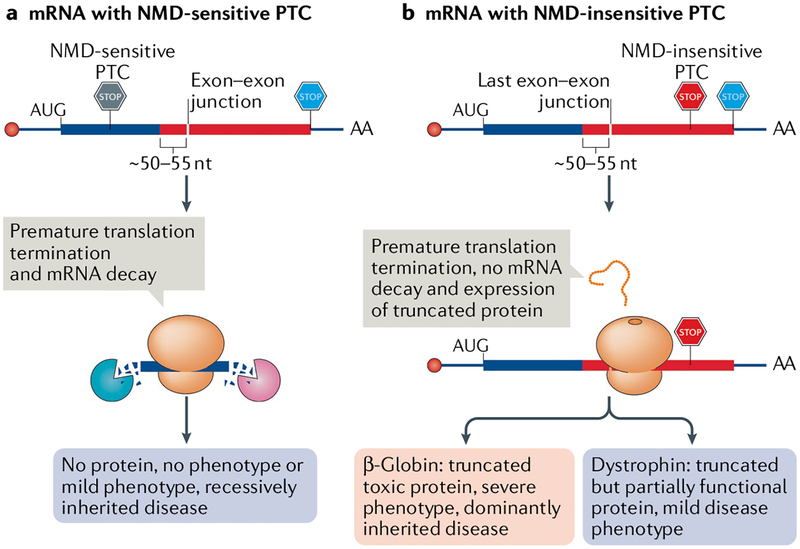
Nonsense-mediated mRNA decay (NMD) is one of the best characterized and most evolutionarily conserved cellular quality control mechanisms. Although NMD was first found to target one-third of mutated, disease-causing mRNAs, it is now known to also target ~10% of unmutated mammalian mRNAs to facilitate appropriate cellular responses - adaptation, differentiation or death - to environmental changes. Mutations in NMD genes in humans are associated with intellectual disability and cancer. In this Review, we discuss how NMD serves multiple purposes in human cells by degrading both mutated mRNAs to protect the integrity of the transcriptome and normal mRNAs to control the quantities of unmutated transcripts.


Conflict of interest statement

Competing interests

The authors declare no competing interests.

Figures

Fig. 1 |
Fig. 1 |. Discriminating between targets and nontargets of nonsense-mediated mRNA decay.
Pre-mRNA splicing is accompanied by deposition of an exon junction complex (EJC) and EJC-associating nonsense-mediated mRNA decay (NMD) factors, such as UPF3X, ~24 nucleotides (nt) upstream of each exon–exon junction. The mRNAs are exported to the cytoplasm and translated. a | 3′ untranslated region (UTR) EJC-mediated NMD. Should translation terminate at a premature termination codon (PTC) that is located ≥50–55 nt upstream of an exon–exon junction, the ribosome will not dislocate the EJC, which is effectively in the 3′ UTR. Because most termination codons are located in the final exon and thus downstream of exon–exon junctions, this situation is abnormal, since an EJC is located downstream of the termination codon. Such translation termination is inefficient, presumably because the EJC interferes with the interaction between polyadenylate-binding protein 1 (PABPC1) and eukaryotic release factor 3 (eRF3). UPF1 and the complex of serine/threonine kinases SMG1–SMG8–SMG9 then join the eRF1–eRF3 translation termination complex and form the SMG1–UPF1–eRFs (SURF) complex. Next, UPF1–SMG1 join the downstream EJC and form the decay-inducing (DECID) complex, where UPF1 is activated by SMG1-mediated phosphorylation (P). UPF1 phosphorylation represses further translation initiation and triggers mRNA decay, which is accomplished by recruiting nucleases, either directly, as in the case of endonucleolytic decay by SMG6, or indirectly (not shown), as in the case of exonucleolytic decay through the SMG5–SMG7 heterodimer. b | EJC-independent NMD. At unusually long, unstructured 3′ UTRs, PABPC1 is too distant from the PTC to efficiently recruit eRF1–eRF3 to initiate translation termination. The presence of UPF1 on the 3′ UTR increases the probability of UPF1 activation by phosphorylation and thus the probability of NMD. c | No NMD. Normally, translating ribosomes remove EJCs and any promiscuously bound UPF1 from the 5′ UTR and from the coding sequence (CDS) but do not travel beyond the stop codon into the 3′ UTR. PABPC1, through its interactions with eRF1–eRF3 or translation initiation factor eIF4G (not shown), is thought to preclude UPF1 from joining the termination complex. CBP, cap-binding protein; eIF4A3, eukaryotic initiation factor 4A3; RBM8A, RNA-binding protein 8A.
Fig. 2 |
Fig. 2 |. UPF1 binding to mRNA and activation of nonsense-mediated mRNA decay.
Cellular UPF1 is largely nonphosphorylated and promiscuously binds to and dissociates from accessible RNA, including mRNAs, using its ATP-dependent 5′-to-3′ RNA helicase activity. UPF1 bound to the mRNA 5′ untranslated region (5′ UTR) and coding sequence is also actively removed by translocating ribosomes, and as a result, UPF1 is relatively enriched on the mRNA 3′ UTR. UPF1 binding to mRNA can activate nonsense-mediated mRNA decay (NMD) through different mechanisms. A 3′ UTR exon junction complex (EJC) increases UPF1 phosphorylation (P) through protein-mediated interactions, whereas a long 3′ UTR (>1 kb) can increase UPF1 occupancy on the 3′ UTR and thus the probability of UPF1 phosphorylation. In both cases, NMD is activated by serine/threonine kinase SMG1-meditated UPF1 phosphorylation, which in turn results in translation repression and recruitment of mRNA decay factors such as SMG5–SMG7 and SMG6. eIF4A3, eukaryotic initiation factor 4A3; eRF, eukaryotic release factor; RBM8A, RNA-binding protein 8A.
Fig. 3 |
Fig. 3 |. Degradation of the nonsense-mediated mRNA decay targets.
Following triggering of nonsense-mediated mRNA decay (NMD) by translation termination through serine/threonine kinase SMG1-mediated UPF1 phosphorylation (P), phosphorylated UPF1 recruits either the endonuclease SMG6 or the SMG5–SMG7 complex. SMG6 cleaves the mRNA near the premature termination codon (PTC), whereas SMG5–SMG7 recruits the deadenylation complex CCR4–NOT through its subunit POP2 and the decapping complex mRNA-decapping enzyme 2 (DCP2)–DCP1a through its subunit PNRC2. DCP2–DCP1a can also be recruited by CCR4–NOT (not shown). SMG5–SMG7 also recruits protein phosphatase 2A (PP2A), which dephosphorylates UPF1. NMD intermediates from either pathway are degraded 5′-to-3′ by the exonuclease XRN1 and 3′-to-5′ by the exosome or the exosome-free 3′-to-5′ exonuclease DIS3-like exonuclease 2 (DIS3L2). Terminal uridylyltransferase 4 (TUT4) and TUT7 can append nontemplated uridines at the 3′ ends; the uridylated decay intermediates are favoured for degradation by DIS3L2. eRF, eukaryotic release factor.
Fig. 4 |
Fig. 4 |. Features of cellular mRNAs that activate nonsense-mediated mRNA decay.
a | An exon–exon junction located ≥50–55 nucleotides (nt) downstream of either a premature termination codon (PTC) or a normal termination codon, with an exon junction complex (EJC) deposited ~24 nt upstream of the junction so that it is far enough from the termination codon and cannot be removed by the terminating ribosome. b | A long (more than ~1 kb), unstructured 3′ untranslated region (3′ UTR), which places the nonsense-mediated mRNA decay (NMD) suppressor polyadenylate-binding protein 1 (PABPC1) too far from the termination codon. c | An upstream open reading frame (uORF) whose termination codon is interpreted as a PTC because an EJC lies ≥50–55 nt downstream of it. d | Alternative splicing (AS) that generates a termination codon that can function as a PTC with any of the above-described features. Alternative splicing may also convert a normal termination codon into one that triggers NMD. e | Alternative 3′-end formation that generates either a 3′ UTR EJC (not shown) or a long 3′ UTR. f | In the few selenoprotein-encoding mRNAs, a UGA codon encoding selenocysteine in cis to a selenocysteine insertion element in a 3′ UTR. The selenocysteine insertion element normally directs the incorporation of selenocysteine, but when cellular selenocysteine concentrations are low, the process becomes inefficient and the UGA codon may be read as a termination codon, which, if properly placed, leads to NMD. eIF4A3, eukaryotic initiation factor 4A3; RBM8A, RNA-binding protein 8A.
Fig. 5 |
Fig. 5 |. Physiological roles of nonsense-mediated mRNA decay.
a | Various extracellular stresses can induce the integrated stress response (ISR). During the ISR, eukaryotic initiation factor 2α (eIF2α) is phosphorylated (P), leading to the suppression of nonsense-mediated mRNA decay (NMD). NMD suppression enables the expression of NMD targets, including those encoding the transcription factors activating transcription factor 4 (ATF4), CCAAT-enhancer-binding protein homologous protein (CHOP) and ATF3, which coordinate the expression of proteins that alleviate the stresses. Following resolution of the ISR, NMD is resumed and suppresses the expression of ATF4 and CHOP, because their mRNAs contain an upstream open reading frame (uORF), and of ATF3, because of a premature termination codon introduced by alternative splicing (AS), thereby ensuring that the ISR is only active during stress. b | Cells that constitutively express the oncogene MYC produce toxic reactive oxygen species (ROS), which activate the ISR and cause eIF2α phosphorylation and NMD suppression, thereby stabilizing SLC7A11 mRNA. SLC7A11 encodes a transporter that imports cystine into the cytoplasm, which is used in the synthesis of the ROS scavenging molecule glutathione. c | Cells with increases in protein-folding demand in the endoplasmic reticulum (ER) induce the unfolded protein response (UPR) in order to increase the folding capacity of the ER (left). The UPR inhibits NMD, and as a result, NMD targets, such as the IRE1A mRNA, which has an unusually long 3′ untranslated region (3′ UTR), are stabilized. IRE1α together with the products of many other natural NMD targets then coordinates the UPR. When the protein-folding capacity of the ER matches demand (right), NMD is active and degrades the IRE1A mRNA and other mRNAs encoding key UPR regulators, thereby ensuring that innocuous stresses do not activate the UPR. d | NMD maintains the pluripotency of mouse neural stem cells by targeting Smad7 mRNA, which encodes a negative regulator of TGFβ signalling. During neural differentiation, the brain-specific microRNA miR-128 is expressed and inhibits the exon junction complex (EJC) component metastatic lymph node 51 (MLN51) and UPF1. This suppresses NMD, enables the production of SMAD7 and facilitates differentiation.
Fig. 6 |
Fig. 6 |. Involvement of nonsense-mediated mRNA decay in disease.
Frameshift or nonsense mutations often introduce a nonsense-mediated mRNA decay (NMD)-sensitive or an NMD-insensitive premature termination codon (PTC). a | mRNAs harbouring an NMD-sensitive PTC that is located ≥50–55 nucleotides (nt) upstream of the last exon–exon junction (PTCs located in the thick blue bar) efficiently undergo NMD, which in turn prevents the production of truncated, potentially toxic proteins. As a consequence, these mutations are recessively inherited and cause either no or only a mild disease in heterozygous individuals. b | mRNAs harbouring an NMD-insensitive PTC, which is generally located ≤50–55 nt upstream of the last exon–exon junction (not shown) or in the last exon (located in the thick red bar), fail to trigger NMD and produce truncated proteins. These truncated proteins could be stable and toxic, as in the case of some β-globin gene mutations (pink). Such mutations can be dominant and patients show a severe disease phenotype, even with one normal allele. However, in some diseases, as illustrated by Becker muscular dystrophy (purple), the truncated protein shows some residual activity, thereby causing milder symptoms than seen in Duchenne muscular dystrophy, in which NMD-sensitive PTCs eliminate dystrophin protein expression.

References

    1. Leeds P, Peltz SW, Jacobson A & Culbertson MR The product of the yeast UPF1 gene is required for rapid turnover of mRNAs containing a premature translational termination codon. Genes Dev 5, 2303–2314 (1991). - PubMed
    1. Atkin AL, Altamura N, Leeds P & Culbertson MR The majority of yeast UPF1 co-localizes with polyribosomes in the cytoplasm. Mol. Biol. Cell 6, 611–625 (1995). - PMC - PubMed
    1. Pal M, Ishigaki Y, Nagy E & Maquat LE Evidence that phosphorylation of human Upf1 protein varies with intracellular location and is mediated by a wortmannin-sensitive and rapamycin-sensitive PI 3-kinase-related kinase signaling pathway. RNA 7, 5–15 (2001). - PMC - PubMed
    1. Czaplinski K et al. The surveillance complex interacts with the translation release factors to enhance termination and degrade aberrant mRNAs. Genes Dev 12, 1665–1677 (1998). - PMC - PubMed
    1. Ivanov PV, Gehring NH, Kunz JB, Hentze MW & Kulozik AE Interactions between UPF1, eRFs, PABP and the exon junction complex suggest an integrated model for mammalian NMD pathways. EMBO J 27, 736–747 (2008). - PMC - PubMed

Publication types