Cytology Microarray on Cell Block Preparation: A Novel Diagnostic Approach in Fluid Cytology
- PMID: 30992641
- PMCID: PMC6425782
- DOI: 10.4103/JOC.JOC_15_17
Cytology Microarray on Cell Block Preparation: A Novel Diagnostic Approach in Fluid Cytology
Abstract
Background: The cytological examination of serous body effusions to diagnose and stage malignancy is well accepted in clinical medicine. Conventional smear (CS) and cell block (CB) study has to be complemented with immunohistochemistry (IHC) for a definitive diagnosis of malignancy and also to differentiate it from reactive mesothelial cells. Cytology microarray (CMA) is a modification of tissue microarray which involves core needle biopsy of multiple cell blocks and embedding it in a single block.
Aim: The aim of this study was to assess the effectiveness of IHC technique in CMA for rapid diagnosis of malignancy and to reduce the cost of testing.
Materials and methods: In this study, 82 pleural fluids were collected and subjected to CS and CB study followed by IHC in CMA blocks. Six commonly used antibodies were applied to confirm malignancy and diagnose the primary.
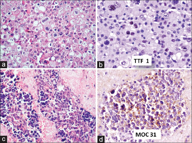
Results: Nineteen cases were diagnosed as malignancy by CB method. MOC-31 confirmed adenocarcinoma deposit in 67% cases of which 44% were proved to be of lung primary by TTF1.
Conclusions: IHC on CMA blocks of effusion fluids is a very effective technique that can significantly reduce the cost of testing by >70%.
Keywords: Cell block; conventional smear; cytology microarray.
Conflict of interest statement
There are no conflicts of interest.
Figures

References
-
- Dekker A, Bupp PA. Cytology of serous effusions. An investigation into the usefulness of cell blocks versus smears. Am J Clin Pathol. 1978;70:855–60. - PubMed
-
- Zhang D, Salto-Tellez M, Putti TC, Do E, Koay ES. Reliability of tissue microarrays in detecting protein expression and gene amplification in breast cancer. Mod Pathol. 2003;16:79–84. - PubMed
-
- Leversha MA, Fielding P, Watson S, Gosney JR, Field JK. Expression of p53, pRB, and p16 in lung tumours: A validation study on tissue microarrays. J Pathol. 2003;200:610–9. - PubMed
-
- Wen CH, Su YC, Wang SL, Yang SF, Chai CY. Application of the microarray technique to cell blocks. Acta Cytol. 2007;51:42–6. - PubMed