

Review
doi: 10.1002/cld.686.
eCollection 2018 Jan.
Imaging features of hepatic arterial and venous flow abnormalities
Affiliations
- PMID: 30992783
- PMCID: PMC6385935
- DOI: 10.1002/cld.686
Item in Clipboard
Review
Imaging features of hepatic arterial and venous flow abnormalities
Clin Liver Dis (Hoboken).
.
No abstract available
Figures
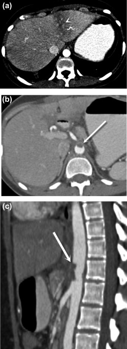
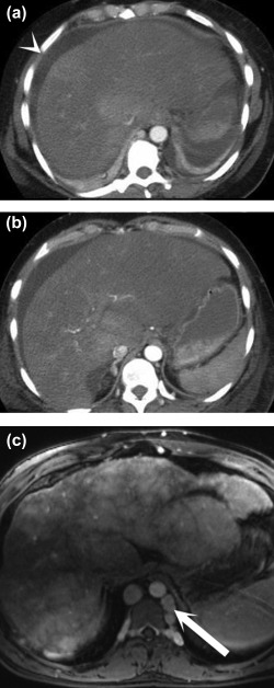
Postoperative aortic and celiac thrombosis. Axial (A, B) and sagittal (C) postcontrast CT images demonstrate a wedge‐shaped area of low attenuation in the left hepatic lobe (A) (arrowhead), representing transient hepatic attenuation difference secondary to hypoperfusion caused by aortic thrombus extending into the celiac trunk, (B, C) (arrows) which formed after endovascular treatment for median arcuate ligament syndrome.
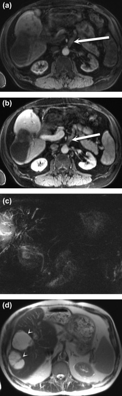
Bile duct necrosis and biloma formation after arterial occlusion complicating liver transplantation. Arterial phase (A) and portal venous phase (B) postcontrast fat‐saturated T1‐weighted MRI sequences show an occluded arterial conduit for transplant (arrows). Biliary necrosis resulted in multiple intrahepatic biliary strictures, seen on the T2‐weighted maximum intensity projection MRCP image (C), and intrahepatic fluid collections consistent with bilomas (arrowheads), seen on the T2‐weighted single‐shot fast spin‐echo MR image (D).
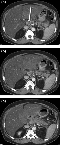
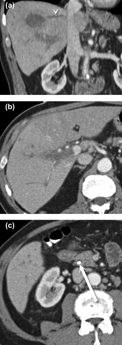
Portal venous thrombosis secondary to pancreatitis. Axial postcontrast CT images (A‐C) demonstrate retroperitoneal fluid and fat stranding surrounding the pancreatic body and tail (C) (arrowhead), consistent with acute pancreatitis. There is associated thrombosis of the splenic vein that extends to the portosplenic confluence, where there is nearly occlusive thrombus in the main portal vein (A) (arrow).
Portal vein tumor thrombus. Axial noncontrast (A) and postcontrast arterial (B) and portal venous (C) phase T1 three‐dimensional gradient‐echo fat‐saturated MR images demonstrate a hypointense lesion in the right lobe of the liver (arrowhead), consistent with hepatocellular carcinoma. An enhancing tumor thrombus is seen in the adjacent portal vein (C) (arrow).
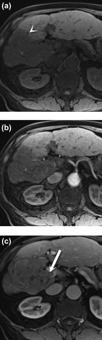
Acute versus chronic Budd‐Chiari syndrome. Postcontrast axial CT images (A and B) show a large, heterogeneous liver, ascites (arrowhead), and nonvisualization of the hepatic veins. Postcontrast T1‐weighted MR image (C) demonstrates the classic “nutmeg” appearance of the parenchyma, surface nodularity (a sign of atrophy), and collateral formation (arrow).
Hepatic vein thrombophlebitis. Coronal (A) and axial (B, C) CT images show an abscess in the dome of the liver (A), which has associated thrombosis of the middle hepatic vein (A) (arrowhead), as well as the right main portal vein (B). The patient improved after treatment for cholangitis arising from an obstructing common bile duct stone (C) (arrow).
Similar articles
-
Hemodynamics of experimental portal venous occlusion in dogs.Am Surg. 1975 Apr;41(4):198-202. Am Surg. 1975. PMID: 1122071
-
Hepatic arterial and portal venous flows during hemorrhage.Eur Surg Res. 1975;7(4-5):259-68. doi: 10.1159/000127811. Eur Surg Res. 1975. PMID: 1181189
-
Role of the hepatic artery in the metabolism of phenacetin and acetaminophen: intravital microscopic and multiple-indicator dilution study in perfused rat liver.Hepatology. 1994 Sep;20(3):672-83. Hepatology. 1994. PMID: 8076925
-
Simultaneous measurement of hepatic arterial and portal venous flows by transit time ultrasonic volume flowmetry.Surg Gynecol Obstet. 1988 Jul;167(1):65-9. Surg Gynecol Obstet. 1988. PMID: 2968000
-
Improved diagnosis of hepatic perfusion disorders: value of hepatic arterial phase imaging during helical CT.Radiographics. 2001 Jan-Feb;21(1):65-81; questionnaire 288-94. doi: 10.1148/radiographics.21.1.g01ja0165. Radiographics. 2001. PMID: 11158645 Review.
Cited by
-
Wandering intravascular air gun BB pellet.Radiol Case Rep. 2020 Oct 12;15(12):2627-2631. doi: 10.1016/j.radcr.2020.09.055. eCollection 2020 Dec. Radiol Case Rep. 2020. PMID: 33088376 Free PMC article.
-
Lessons of the month: Acute liver failure: a case close to the heart.Clin Med (Lond). 2021 Mar;21(2):e234-e236. doi: 10.7861/clinmed.2020-1062. Clin Med (Lond). 2021. PMID: 33762393 Free PMC article.
-
Correlation between portal vein diameter and craniocaudal length of the spleen.J Ultrason. 2019 Dec;19(79):276-281. doi: 10.15557/JoU.2019.0041. Epub 2019 Dec 31. J Ultrason. 2019. PMID: 32021709 Free PMC article.
-
Risk Factors for In-hospital Mortality After Transarterial Intervention After Postpancreatectomy Hemorrhage.Cardiovasc Intervent Radiol. 2020 Sep;43(9):1342-1352. doi: 10.1007/s00270-020-02509-2. Epub 2020 May 20. Cardiovasc Intervent Radiol. 2020. PMID: 32435837 Free PMC article.
-
Eosinophilic Pneumonia Induced by Daptomycin.Cureus. 2024 Feb 27;16(2):e55095. doi: 10.7759/cureus.55095. eCollection 2024 Feb. Cureus. 2024. PMID: 38558746 Free PMC article.
References
-
- Ryan MF, Hamilton PA, Sarrazin J, Chu P, Benjaminov O, Lam K. The halo sign and peripancreatic fluid: useful CT signs of hypovolaemic shock complex in adults. Clin Radiol 2005;60:599‐607. - PubMed
-
- Lubner M, Demertzis J, Lee JY, Appleton CM, Bhalla S, Menias CO. CT evaluation of shock viscera: a pictorial review. Emerg Radiol 2008;15:1‐11. - PubMed
-
- Oderich G, Panneton JM, Bower TC, Ricotta JJ 2nd, Sundt TM 3rd, Cha S, Gloviczki P. Aortic dissection with aortic side branch compromise: impact of malperfusion on patient outcome. Perspect Vasc Surg Endovasc Ther 2008;20:190‐200. - PubMed
-
- Holbert B, Baron RL, Dodd GD 3rd. Hepatic infarction caused by arterial insufficiency. AJR Am J Roentgenol 1996;166:815‐820. - PubMed
-
- Raab BW. The thread and streak sign. Radiology 2005;236:284‐285. - PubMed
Publication types
LinkOut - more resources
Full Text Sources
