

Lymphangiomatosis of the ileum with perforation: A case report and review of the literature
- PMID: 30992989
- PMCID: PMC6449703
- DOI: 10.1016/j.amsu.2019.03.010
Lymphangiomatosis of the ileum with perforation: A case report and review of the literature
Abstract
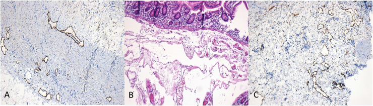
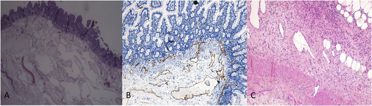
Lymphangiomatosis is a benign proliferation of lymph vessels. Lymphatic diseases can vary from small lymphangioma to generalized lymphangiomatosis, which is a rare condition and can have several clinical manifestations. The gastrointestinal tract may be affected, but the incidence in the intestinal wall is very low. We propose in our study a case of ileal lymphangiomatosis presenting with perforation, in which the diagnosis was made after the pathological analysis of the resected intestinal tract. Although rare and not described in the literature, intestinal lymphangiomatosis could manifest itself with acute abdomen and could be a surgical urgency. This disease should be considered when intestinal perforation is observed.
Keywords: Acute abdomen; Bowel perforation; Bowel resection; Lymphangiomatosis; Small intestine.
Figures
References
-
- Valakada J., Madhusudhan K.S., Ranjan g. Abdominal lymphangiomatosis with intestinal lymphangiectasia diagnosed by magnetic resonance lymphangiography: a case report. Curr. Probl. Diagn. Radiol. 2018 May - Jun;47(3):200–202. - PubMed
-
- Puri A.S., Aggarwal R., Gupta R.K. Intestinal lymphangiectasia: evaluation by CT and scintigraphy. Gastrointest. Radiol. 1992;17(2):119–121. - PubMed
Publication types
LinkOut - more resources
Full Text Sources
