. 2019 Apr 8:7:e6707.
doi: 10.7717/peerj.6707. eCollection 2019.

The unfolding of iRFP713 in a crowded milieu

Affiliations

The unfolding of iRFP713 in a crowded milieu

Olesya V Stepanenko et al. PeerJ. .

Abstract

The exploring of biological processes in vitro under conditions of macromolecular crowding is a way to achieve an understanding of how these processes occur in vivo. In this work, we study the unfolding of the fluorescent probe iRFP713 in crowded environment in vitro. Previously, we showed that the unfolding of the dimeric iRFP713 is accompanied by the formation of a compact monomer and an intermediate state of the protein. In the intermediate state, the macromolecules of iRFP713 have hydrophobic clusters exposed to the surface of the protein and are prone to aggregation. Concentrated solutions of polyethylene glycol (PEG-8000), Dextran-40 and Dextran-70 with a molecular mass of 8000, 40000 and 70000 Da, respectively, were used to model the conditions for macromolecular crowding. A limited available space provided by all the crowding agents used favors to the enhanced aggregation of iRFP713 in the intermediate state at the concentration of guanidine hydrochloride (GdnHCl), at which the charge of protein surface is neutralized by the guanidine cations. This is in line with the theory of the excluded volume. In concentrated solutions of the crowding agents (240-300 mg/ml), the stabilization of the structure of iRFP713 in the intermediate state is observed. PEG-8000 also enhances the stability of iRFP713 in the monomeric compact state, whereas in concentrated solutions of Dextran-40 and Dextran-70 the resistance of the protein in the monomeric state against GdnHCl-induced unfolding decreases. The obtained data argues for the excluded volume effect being not the only factor that contributes the behavior of biological molecules in a crowded milieu. Crowding agents do not affect the structure of the native dimer of iRFP713, which excludes the direct interactions between the target protein and the crowding agents. PEGs of different molecular mass and Dextran-40/Dextran-70 are known to influence the solvent properties of water. The solvent dipolarity/polarizability and basicity/acidity in aqueous solutions of these crowding agents vary in different ways. The change of the solvent properties in aqueous solutions of crowding agents might impact the functioning of a target protein.

Keywords: Crowded milieu; Excluded volume effect; Protein aggregation; Protein unfolding; Solvent properties; iRFP713.


Conflict of interest statement

Konstantin K. Turoverov and Irina M. Kuznetsova are Academic Editors for PeerJ.

Figures

Figure 1
Figure 1. The effect of crowding agent PEG-8000 on the spectral properties of iRFP713 in the holoform.
(A) Absorption spectra, tryptophan fluorescence spectra (λex = 295 nm) and the chromophore fluorescence spectra (λex = 690 nm). (B) CD spectra in the far-UV, near-UV and visible region of the spectrum. The color of the curves corresponds to different concentration of PEG-8000: 0 mg/ml (black line), 80 mg/ml (red line), 120 mg/ml (gray line) and 300 mg/ml (blue line). Absorption and fluorescence spectra are drawn by dashed and solid lines, respectively.
Figure 2
Figure 2. The effect of crowding agents Dextran-40 and Dextran-70 on the spectral properties of iRFP713 in the holoform.
The designations on panels A–B are the same as in the caption to Fig. 1. The color of the curves symbolizes the different experimental conditions: 0 mg/ml crowding agent (black line), 240 mg/ml of Dextran-40 (red line) and 240 mg/ml of Dextran-70 (green line). Absorption and fluorescence spectra are drawn by dashed and solid lines, respectively.
Figure 3
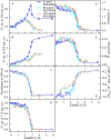
Figure 3. Unfolding of iRFP713 in the holoform induced by guanidine hydrochloride (GdnHCl) in the presence of PEG-8000.
(A and B) Changes in the tryptophan fluorescence intensity at registration wavelengths of 320 and 365 nm. The fluorescence was excited at wavelength of 295 nm. The values of fluorescence intensity I320 and I365 were normalized to unity at zero denaturant concentration. (C) Changes in optical density of the solution; λ = 690 nm. (D) Changes in the chromophore fluorescence intensity at an excitation wavelength of 690 nm, corrected for the primary inner filter effect taking into account changes in the absorbance of the solution at the excitation wavelength (see Materials and Methods). (E) Changes in the parameter A = I320I365 at an excitation wavelength of 295 nm. (F) Changes in fluorescence anisotropy at excitation and emission wavelengths of 295 and 365 nm. (G) Changes in the ellipticity at 222 nm. The color of the curves corresponds to different concentration of PEG-8000: 0 mg/ml (blue circles), 80 mg/ml (cyan circles), 120 mg/ml (gray line) and 300 mg/ml (orange circles). The measurements were performed after 24 h incubation of the native protein in the presence of GdnHCl.
Figure 4
Figure 4. The change in the recorded absorption spectra of iRFP713 in the holoform at GdnHCl-induced unfolding in the presence of PEG-8000 at a concentration of 80 mg/ml.
Numerals at the curves are the final concentration of the denaturant in the protein solutions.
Figure 5
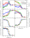
Figure 5. Unfolding of iRFP713 and its mutant variants in their holoforms induced by guanidine hydrochloride (GdnHCl).
The designations for A–F are the same as in the caption to Fig. 3. Symbols of different color correspond to different proteins: iRFP713 (black symbols), iRFP713-W109 (red symbols), iRFP713-W281 (green symbols) and iRFP713-W311 (blue symbols). The measurements were performed after 24 h incubation of proteins in the presence of GdnHCl.
Figure 6
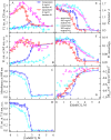
Figure 6. Unfolding of iRFP713 in the apo- and holoform induced by guanidine thiocyanate (GTC) in the presence of PEG-8000 at a concentration of 300 mg/ml.
The designations for A–F are the same as in the caption to Fig. 3. The values of fluorescence intensity I320 and I365 of the holoprotein were normalized to unity at zero denaturant concentration. The values of fluorescence intensity I320 and I365 of the apoprotein were normalized to a value that is equal to the ratio of the fluorescence intensity at the corresponding registration wavelength of apo- and holoprotein at zero denaturant concentration. The color of the symbols indicates the concentration of PEG-8000 in solutions of apo- and holoprotein: 0 mg/ml (red and blue circles, respectively), 300 mg/ml (pink and cyan circles, respectively). The measurements were performed after 24 h incubation of apo- or holoprotein in the presence of GdnHCl.
Figure 7
Figure 7. Unfolding of iRFP713 in the apo- and holoform induced by guanidine hydrochloride (GdnHCl) in the presence of Dextran-70.
The designations for A–G are the same as in the caption to Fig. 3. The color of the symbols denotes the concentration of dextran-70 in solutions of apo- and holoprotein: 0 mg/ml (red and blue circles, respectively), 240 mg/ml (pink and cyan circles, respectively). The measurements were performed after 24 h incubation of apo- or holoprotein in the presence of GdnHCl.
Figure 8
Figure 8. Unfolding of iRFP713 in the apo- and holoform induced by guanidine hydrochloride (GdnHCl) in the presence of Dextran-40.
The designations for A–G are the same as in the caption to Fig. 3. The color of the symbols denotes the concentration of dextran-40 in solutions of apo- and holoprotein: 0 mg/ml (red and blue circles, respectively), 240 mg/ml (pink and cyan circles, respectively). The measurements were performed after 24 h incubation of apo- or holoprotein in the presence of GdnHCl.
Figure 9
Figure 9. The change in the recorded absorption spectra of iRFP713 in the holoform at GdnHCl-induced unfolding in the presence of Dextran-40 at a concentration of 240 mg/ml.
Numerals at the curves show the final concentration of the denaturant in the protein solutions.

Similar articles

References

    1. Al-Ayoubi SR, Schummel PH, Golub M, Peters J, Winter R. Influence of cosolvents, self-crowding, temperature and pressure on the sub-nanosecond dynamics and folding stability of lysozyme. Physical Chemistry Chemical Physics. 2017;19:14230–14237. doi: 10.1039/C7CP00705A. - DOI - PubMed
    1. Bai J, Liu M, Pielak GJ, Li C. Macromolecular and small molecular crowding have similar effects on alpha-synuclein structure. Chemphyschem. 2017;18:55–58. doi: 10.1002/cphc.201601097. - DOI - PubMed
    1. Bhoo SH, Davis SJ, Walker J, Karniol B, Vierstra RD. Bacteriophytochromes are photochromic histidine kinases using a biliverdin chromophore. Nature. 2001;414:776–779. doi: 10.1038/414776a. - DOI - PubMed
    1. Chebotareva NA, Filippov DO, Kurganov BI. Effect of crowding on several stages of protein aggregation in test systems in the presence of alpha-crystallin. International Journal of Biological Macromolecules. 2015;80:358–365. doi: 10.1016/j.ijbiomac.2015.07.002. - DOI - PubMed
    1. Chen M, Li W, Zhang Z, Liu S, Zhang X, Zhang XE, Cui Z. Novel near-infrared BiFC systems from a bacterial phytochrome for imaging protein interactions and drug evaluation under physiological conditions. Biomaterials. 2015;48:97–107. doi: 10.1016/j.biomaterials.2015.01.038. - DOI - PubMed

LinkOut - more resources