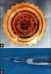
Step-by-step Descemet's membrane endothelial keratoplasty surgery
- PMID: 30993063
- PMCID: PMC6432849
- DOI: 10.4103/tjo.tjo_108_18
Step-by-step Descemet's membrane endothelial keratoplasty surgery
Abstract
With the success of Descemet's stripping automated endothelial keratoplasty (DSAEK) technique providing better outcomes in visual prognosis and theoretically lower rejection rate than penetrating keratoplasty, DSAEK dominated the realm of endothelial keratoplasty for the past decade. However, Descemet's membrane endothelial keratoplasty (DMEK) has become more and more popular worldwide due to its even lower rejection rate, faster visual recovery, better visual outcome, and lower long-term endothelial loss. In this article, we demonstrate the techniques and nuances of DMEK surgery in detail for corneal specialists who are beginning their DMEK surgeries.
Keywords: Corneal transplantation; Descemet membrane endothelial keratoplasty; endothelial keratoplasty.
Conflict of interest statement
The authors declare that there are no conflicts of interests of this paper.
Figures


References
-
- Zirm EK. Eine erfolgreiche totale keratoplastik (A successful total keratoplasty).1906. Refract Corneal Surg. 1989;5:258–61. - PubMed
-
- Tillett CW. Posterior lamellar keratoplasty. Am J Ophthalmol. 1956;41:530–3. - PubMed
-
- Ko WW, Frueh BE, Shields CK, Costello ML, Feldman ST. Experimental posterior lamellar transplantation of the rabbit cornea. (ARVO abstract) Invest Ophthalmol Vis Sci. 1993;34:1102.
-
- Terry MA, Ousley PJ. Deep lamellar endothelial keratoplasty in the first United States patients: Early clinical results. Cornea. 2001;20:239–43. - PubMed
-
- Nieuwendaal CP, Lapid-Gortzak R, van der Meulen IJ, Melles GJ. Posterior lamellar keratoplasty using descemetorhexis and organ-cultured donor corneal tissue (Melles technique) Cornea. 2006;25:933–6. - PubMed
Publication types
LinkOut - more resources
Full Text Sources
Miscellaneous

