. 2019 Apr 15:3:11.
doi: 10.1038/s41698-019-0083-4. eCollection 2019.

Precise definition of PTEN C-terminal epitopes and its implications in clinical oncology

Affiliations

Precise definition of PTEN C-terminal epitopes and its implications in clinical oncology

Janire Mingo et al. NPJ Precis Oncol. .

Abstract

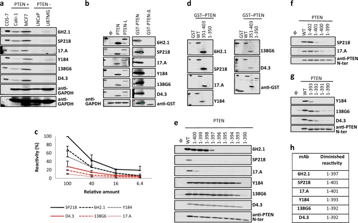
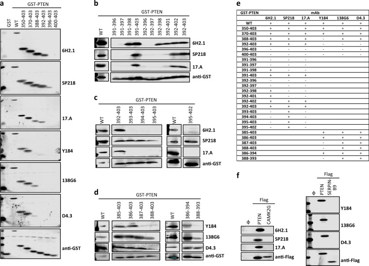
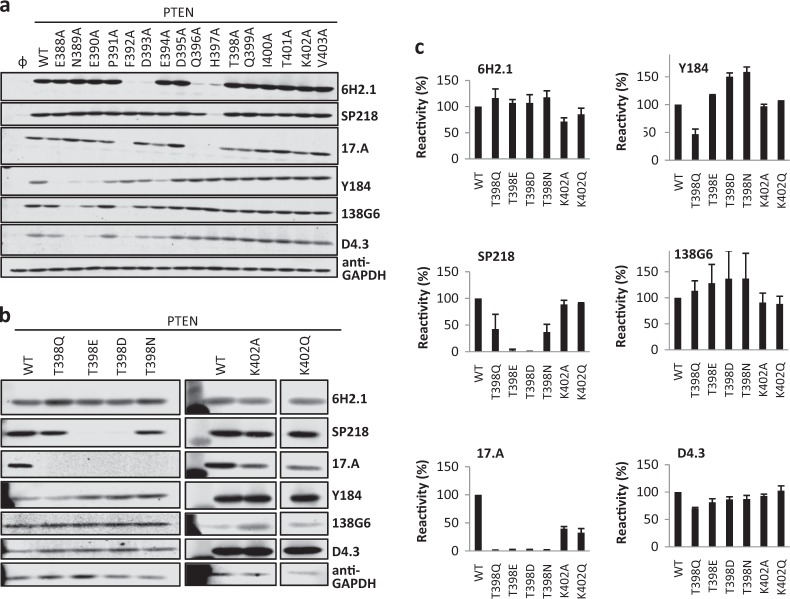
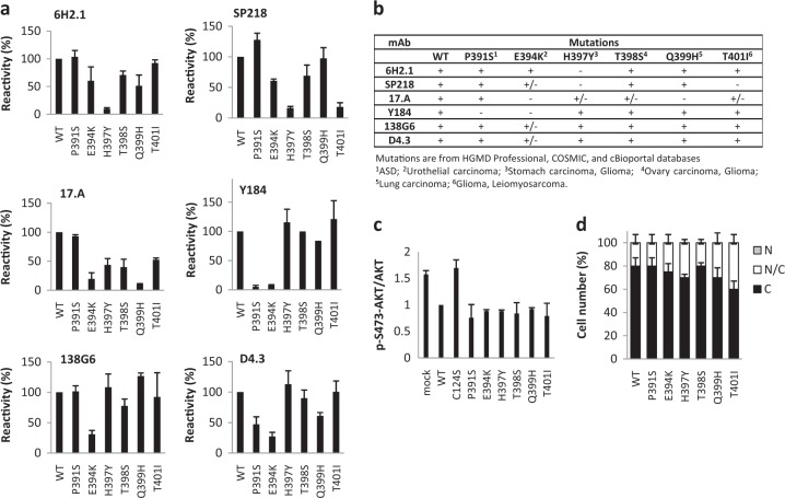
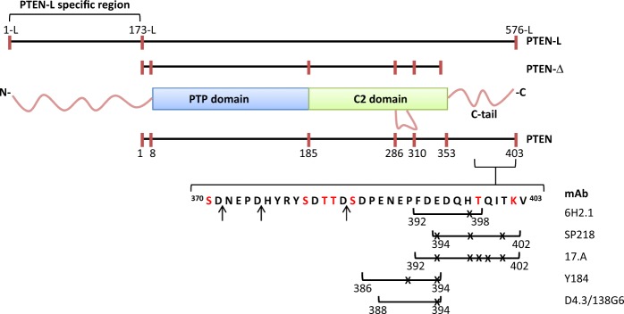
Anti-PTEN monoclonal antibodies (mAb) are arising as important tools for immunohistochemistry (IHC) and protein quantification routine analysis in clinical oncology. Although an effort has been made to document the reliability of tumor tissue section immunostaining by anti-PTEN mAb, and to standardize their IHC use in research and in the clinical practice, the precise topological and biochemical definition of the epitope recognized by each mAb has been conventionally overlooked. In this study, six commercial anti-PTEN mAb have been validated and characterized for sensitivity and specificity by IHC and FISH, using a set of prostate and urothelial bladder tumor specimens, and by immunoblot, using PTEN positive and PTEN negative human cell lines. Immunoblot precise epitope mapping, performed using recombinant PTEN variants and mutations, revealed that all mAb recognized linear epitopes of 6-11 amino acid length at the PTEN C-terminus. Tumor-associated or disease-associated mutations at the PTEN C-terminus did not affect subcellular localization or PIP3 phosphatase activity of PTEN in cells, although resulted in specific loss of reactivity for some mAb. Furthermore, specific mimicking-phosphorylation mutations at the PTEN C-terminal region also abolished binding of specific mAb. Our study adds new evidence on the relevance of a precise epitope mapping in the validation of anti-PTEN mAb for their use in the clinics. This will be substantial to provide a more accurate diagnosis in clinical oncology based on PTEN protein expression in tumors and biological fluids.


Conflict of interest statement

The authors declare no competing interests.

Figures

Fig. 1
Fig. 1
Specificity, sensitivity, and reactivity of anti-PTEN mAb with PTEN C-terminus. a Specificity of anti-PTEN mAb. Detection of endogenous PTEN protein by the different anti-PTEN mAb by immunoblot, using cell lysates from PTEN-positive (Caki-1 and MCF7) and PTEN-negative (LNCaP and U87MG) cell lines. Low-exposure and high-exposure images of anti-GAPDH blots are shown as a control. Detection of recombinant PTEN 1–403, from lysates from transfected COS-7 cells, is also shown. b Recognition of PTEN isoforms. In the left panel, detection of recombinant PTEN 1–403 and PTEN-L 1-L-576-L by immunoblot from lysates from transfected COS-7 cells is shown, and detection of GAPDH with anti-GAPDH antibody is shown as a control. In the right panel, detection of recombinant GST-PTEN 1-403 and GST-PTEN-Δ 1-343-Ser is shown, and detection using anti-GST antibody is shown as a control. Ø, empty vector. c Sensitivity of anti-PTEN mAb. Reactivity of the anti-PTEN mAb with decreasing amounts of recombinant PTEN from cell lysates from transfected COS-7 cells. Data are shown as relative mAb reactivity by immunoblot (mean ± s.d. from three independent experiments), as determined by PTEN protein band quantification. d Reactivity of anti-PTEN mAb with PTEN C-terminus. Detection of GST PTEN 1–403 (WT), GST PTEN 351–403, and GST PTEN 1–350 by the different anti-PTEN mAb by immunoblot, as in b. Detection using anti-GST antibody is also shown as a control. GST, GST alone. eg Reactivity of anti-PTEN mAb with PTEN C-terminus. Detection of PTEN C-terminal truncations, as in b. Detection using a polyclonal antibody recognizing PTEN N-terminus (anti-PTEN N-ter) is also shown as a control. WT, PTEN 1-403; Ø, empty vector. h Summary of the diminished reactivity of the different anti-PTEN mAb with PTEN C-terminal truncations
Fig. 2
Fig. 2
Reactivity of anti-PTEN mAb with peptides from PTEN C-terminal tail and with potential cross-reacting proteins. ad Detection of recombinant GST-fusion proteins containing peptides from the PTEN C-terminal tail, as in Fig. 1. Detection using anti-GST antibody is also shown as a control. WT, GST-PTEN 1–403; GST, GST alone. e Summary of the reactivity of the different anti-PTEN mAb with the GST-fusion proteins containing peptides from the PTEN C-terminal tail. +, binding; −, no binding. f Reactivity of the anti-PTEN mAb with SERPINB9-Flag or CAMK2G-Flag. Detection using anti-Flag antibody is shown as a control. Ø, empty vector
Fig. 3
Fig. 3
Reactivity of anti-PTEN mAb with PTEN variants containing amino acid substitutions at PTEN C-terminal tail. a Reactivity of anti-PTEN mAb with Ala-substitution mutations at PTEN C-terminus, as in Fig. 1. Detection of GAPDH with anti-GAPDH antibody is shown as a control. Ø, empty vector. b, c Reactivity of anti-PTEN mAb with amino acid substitutions mimicking phosphorylation or acetylation at PTEN C-terminus, as in Fig. 1. Both immunoblot images (b) and quantification of the relative mAb reactivity (c) (mean ± s.d. from two independent experiments) is shown
Fig. 4
Fig. 4
Recognition by anti-PTEN mAb and functional properties of disease-associated PTEN C-terminal tail variants. a Reactivity of anti-PTEN mAb with amino acid substitutions at PTEN C-terminus found in tumors or in patients. Quantification of the reactivity is shown (mean ± s.d. from two independent experiments), as in Fig. 3c. b Summary of the reactivity of the different anti-PTEN mAb with amino acid substitutions found in tumors or in patients mutations. +, binding; +/− diminished binding; -, no binding. c Functional activity of the disease-associated PTEN variants. Data are shown as the ratio pSer473-AKT/AKT (mean ± s.d. from two independent experiments), quantified by immunoblot with anti-pSer473-AKT and anti-AKT antibodies, from lysates from COS-7 cells co-transfected with PTEN and AKT1. d Subcellular localization of disease-associated PTEN variants. Data are shown as the percentage of cells displaying cytoplasmic (C), nuclear/cytoplasmic (N/C), or nuclear (N) localization (mean ± s.d. from two independent experiments)
Fig. 5
Fig. 5
Schematic depiction of PTEN isoforms indicating the amino acid sequences of the linear epitopes recognized by the different anti-PTEN mAb. Numbers indicate the amino acid numbers. Numbering of PTEN-L is according to ref. Amino acids are indicated by one-letter code. Ser and Thr residues in red are physiologically phosphorylated residues. The Lys residue in red can undergo acetylation. The arrows indicate the residues where PTEN is cleaved by caspase-3. The lines under the depicted amino acid sequence delimit the epitopes recognized by each mAb. Crosses indicate residues mutated in tumors or in patients whose substitution causes total or partial loss of mAb reactivity

References

    1. Djuric U, Zadeh G, Aldape K, Diamandis P. Precision histology: how deep learning is poised to revitalize histomorphology for personalized cancer care. NPJ Precis. Oncol. 2017;1:22. doi: 10.1038/s41698-017-0022-1. - DOI - PMC - PubMed
    1. Leong TY, Cooper K, Leong AS. Immunohistology—past, present, and future. Adv. Anat. Pathol. 2010;17:404–418. doi: 10.1097/PAP.0b013e3181f8957c. - DOI - PubMed
    1. Schmidt KT, Chau CH, Price DK, Figg WD. Precision oncology medicine: the clinical relevance of patient-specific biomarkers used to optimize cancer treatment. J. Clin. Pharmacol. 2016;56:1484–1499. doi: 10.1002/jcph.765. - DOI - PMC - PubMed
    1. Twomey JD, Brahme NN, Zhang B. Drug-biomarker co-development in oncology—20 years and counting. Drug Resist. Updates. 2017;30:48–62. doi: 10.1016/j.drup.2017.02.002. - DOI - PubMed
    1. Howat WJ, et al. Antibody validation of immunohistochemistry for biomarker discovery: recommendations of a consortium of academic and pharmaceutical based histopathology researchers. Methods. 2014;70:34–38. doi: 10.1016/j.ymeth.2014.01.018. - DOI - PMC - PubMed