. 2019 Oct;76(19):3899-3914.
doi: 10.1007/s00018-019-03102-8. Epub 2019 Apr 16.

Characterization of PPIB interaction in the P3H1 ternary complex and implications for its pathological mutations

Affiliations

Characterization of PPIB interaction in the P3H1 ternary complex and implications for its pathological mutations

Jiawei Wu et al. Cell Mol Life Sci. 2019 Oct.

Abstract

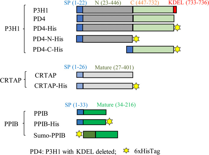
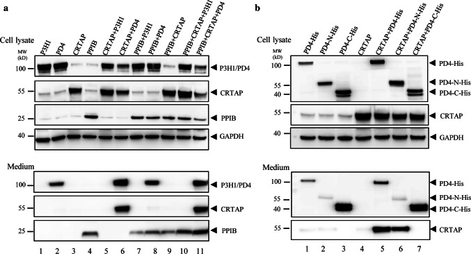
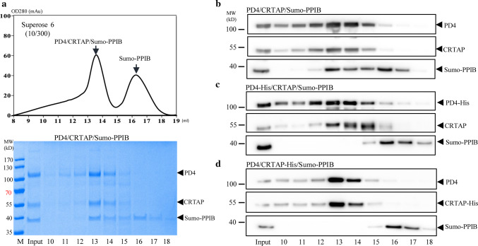
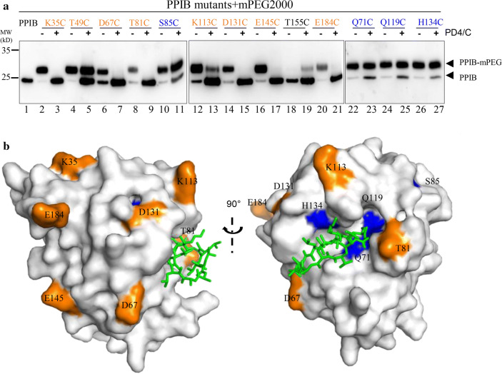
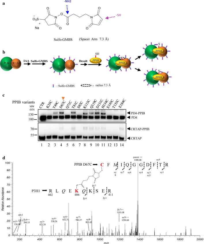
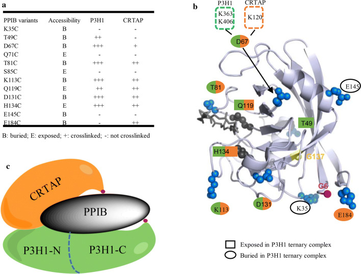
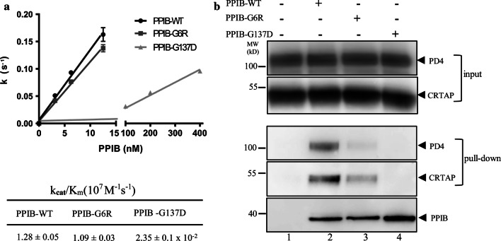
The P3H1/CRTAP/PPIB complex is essential for prolyl 3-hydroxylation and folding of procollagens in the endoplasmic reticulum (ER). Deficiency in any component of this ternary complex is associated with the misfolding of collagen and the onset of osteogenesis imperfecta. However, little structure information is available about how this ternary complex is assembled and retained in the ER. Here, we assessed the role of the KDEL sequence of P3H1 and probed the spatial interactions of PPIB in the complex. We show that the KDEL sequence is essential for retaining the P3H1 complex in the ER. Its removal resulted in co-secretion of P3H1 and CRTAP out of the cell, which was mediated by the binding of P3H1 N-terminal domain with CRTAP. The secreted P3H1/CRTAP can readily bind PPIB with their C-termini close to PPIB in the ternary complex. Cysteine modification, crosslinking, and mass spectrometry experiments identified PPIB surface residues involved in the complex formation, and showed that the surface of PPIB is extensively covered by the binding of P3H1 and CRTAP. Most importantly, we demonstrated that one disease-associated pathological PPIB mutation on the binding interface did not affect the PPIB prolyl-isomerase activity, but disrupted the formation of P3H1/CRTAP/PPIB ternary complex. This suggests that defects in the integrity of the P3H1 ternary complex are associated with pathological collagen misfolding. Taken together, these results provide novel structural information on how PPIB interacts with other components of the P3H1 complex and indicate that the integrity of P3H1 complex is required for proper collagen formation.

Keywords: Chaperone; Hydroxylase; Hyperelastosis cutis; Osteogenesis imperfecta; Sulfo-GMBS.


Figures

Fig. 1
Fig. 1
Schematic representation of P3H1, CRTAP, and PPIB variants. Various constructs were prepared to explore the interactions among P3H1/CRTAP/PPIB complex members. Blue squares represent the signal peptides of each construct, red squares represent the KDEL sequence of P3H1, and the yellow stars represent the His6 sequence. P3H1 with the KDEL sequence removed from the tail was named PD4. The N-terminal segment of P3H1 (1–446) was named PD4-N-His, and the C-terminal dioxygenase segment (447–732) was named PD4-C-His. Sumo-PPIB variant was expressed in E. coli, and all other P3H1, CRTAP, and PPIB variants were prepared from HEK 293E cells
Fig. 2
Fig. 2
The role of KDEL in co-translocation of P3H1/CRTAP. a Transfection of various combinations among P3H1, PD4, CRTAP, and PPIB into cells, followed by the analysis of the media and cell lysates by western blot. Wild-type P3H1 and CRTAP were almost all retained in the cells in HEK 293E cell lines (lanes 1, 3, 5, and 10). Substantial amount of the PD4 was secreted into the medium (lane 2), while large amount of CRTAP was secreted along with PD4 (lanes 6 and 11). This shows the importance of the KDEL sequence in the retention of both P3H1 and CRTAP in ER. GAPDH in the cells was blotted as a loading control. b The full-length of PD4-His, the N-terminal segment of PD4 (PD4-N-His), and the C-terminal dioxygenase domain of PD4 (PD4-C-His) were transfected alone or with CRTAP. The samples were analyzed by SDS-PAGE and western blot. Both domains of P3H1 could be secreted into the culture medium, and CRTAP was mostly retained in the cells; however, when co-expressed with PD4-His (lane 5) or PD4-N-His (lane 6), substantial amount of CRTAP was secreted into the medium. Little CRTAP was detected in the medium when it was co-expressed with PD4-C-His (lane 7). All experiments were performed ≥ 3 times independently
Fig. 3
Fig. 3
The role of the C-termini of P3H1 and CRTAP in forming a complex with PPIB. a Partially purified PD4/CRTAP was mixed with Sumo-PPIB and then analyzed by a Superose 6 gel filtration column with eluted fractions analyzed by coomassie-stained SDS-PAGE. This shows that PPIB can readily form ternary complex with the secreted PD4/CRTAP and was eluted in the early peak with PD4/CRTAP. bd The partially purified PD4/CRTAP or PD4-His/CRTAP or PD4/CRTAP-His were mixed with Sumo-PPIB and then directly analyzed by gel filtration. The elution positions of PD4, CRTAP, and Sumo-PPIB were analyzed by SDS-PAGE and western blot. Complex formation in fraction 13 and 14 was observed from PD4/CRTAP/Sumo-PPIB in b, as seen in a, but not from the mixture of Sumo-PPIB with PD4-His/CRTAP (c) or PD4/CRTAP-His (d). All experiments were performed ≥ 3 times independently
Fig. 4
Fig. 4
Probing the surface area of PPIB that interacts with the PD4/CRTAP complex. a PPIB variants with residues Lys35, Thr49, Asp67, Gln71, Thr81, Ser85, Lys113, Gln119, Asp131, His134, Glu145, Thr155, and Glu184 mutated to cysteine individually, were mixed with mPEG in the absence or presence of the co-expressed PD4/CRTAP. The samples were then analyzed by western blot. mPEG is a cysteine-specific alkylation reagent with each modification of PPIB (PPIB-mPEG), resulting in an increase in the molecular weight of 2000 Da. PPIB variants (S85C, Q71C, Q119C, and H134C), where their modification is not affected by the presence of PD4/CRTAP, are labeled in blue. PPIB variants (K35C, D67C, T81C, K113C, D131C, E145C, and E184C), where their modification is inhibited by the presence of PD4/CRTAP, are labeled in orange. T49C and K113C could be partially modified by mPEG in the presence of PD4 and CRTAP. T155C is resistant to the mPEG modification. b The positions of selected residues on the surface of PPIB structure are colored according to their accessibility in the P3H1 ternary complex. PPIB inhibitor CsA binds PPIB in the substrate-binding pocket and is shown in sticks (green). T49C is located in the backside of the current viewpoint
Fig. 5
Fig. 5
Chemical crosslinking of PPIB variants to the PD4/CRTAP complex. a Sulfo-GMBS is a water-soluble amine-to-sulfhydryl crosslinker with a short spacer arm (7.3 Å). b Sulfo-GMBS conjugates with the free amines of PD4 and CRTAP through its N-hydroxysuccinimide ester. Once PPIB is added, the maleimide group of sulfo-GMBS will form a covalent linkage with a nearby cysteine residue of PPIB variant. c The PD4/CRTAP were incubated with Sulfo-GMBS, and then mixed with the PPIB variants according to the procedure in b, and samples were analyzed by western blot. The high-molecular-weight species represent the crosslinked PD4–PPIB or CRTAP–PPIB complex. The crosslinking data are summarized in Fig. 6a according to the relative density of the bands. d The crosslinked sample of PD4/CRTAP with PPIB D67C was digested with trypsin and analyzed by LC-mass spectrometry with Sulfo-GMBS containing peptides identified. The PPIB D67C was found to crosslink with K406 of P3H1 or K363 of PD4 or K120 of CRTAP (Supplemental Fig. 6)
Fig. 6
Fig. 6
Summarized PPIB intramolecular interactions in the P3H1 ternary complex. a Accessibility of PPIB surface residues in the P3H1 ternary complex was derived from Fig. 4, and the crosslinking information was derived from Fig. 5c. b The key residues of PPIB tested here are highlighted on the surface of PPIB structure showing their accessibility and crosslinking property. The buried PPIB residues upon ternary complex formation are shown in blue spheres. The residues crosslinked to P3H1 (T49, D67, T81, K113, Q119, D131, and H134) are marked in green, and the residues crosslinked to CRTAP (D67, T81, K113, Q119, D131, H134, and E184) are marked in orange. D67 was identified to be close to K363 and K406 (green dotted box) of P3H1, and the K120 (orange dotted box) of CRTAP. c A working model showing relative spatial positions of each component in the P3H1/CRTAP/PPIB complex. The C-termini of P3H1 and CRTAP are labeled as red dots
Fig. 7
Fig. 7
Characterization of pathological PPIB mutants. a The kinetics of PPIB variants in cis/trans isomerization of the Suc-AAPF-pNA substrate were measured by following the absorbance at 390 nm in a spectrophotometer at 8 °C with spectra shown (Supplemental Fig. 7). The rate of cistrans transition in the substrate was plotted against the concentrations of PPIB variants with the calculated kcat/Km values listed at the bottom. The activity of PPIB-G137D is very low and little activity could be detected when its concentration is below 20 nM. The break in the x-axis is between 15 and 100 nM. PPIB-G6R has similar catalyzing activity as the wild-type PPIB; however, the PPIB-G137D mutant is about 45 times less active. b The binding of PPIB variants towards P3H1 and CRTAP was assessed by a pull-down assay. PPIB variants immobilized on Ni magnetic beads were incubated with partially purified PD4/CRTAP. The bound proteins were then analyzed by SDS-PAGE and western blot using antibodies against P3H1 and CRTAP, respectively. Lane 1, immobilized Sumo protein as a control; lane 2, Sumo-PPIB-WT; lane 3, Sumo-PPIB G6R; lane 4, Sumo-PPIB G137D. The two pathological mutations G6R and G137D largely abolished the ability of PPIB to form ternary complexes with P3H1 and CRTAP (lane 3, 4). All experiments were performed ≥ 3 times independently. Error bars represent mean ± SEM

Similar articles

Cited by

References

    1. Bächinger HP, Mizuno K, Vranka JA, Boudko SP. Collagen formation and structure. In: Mander L, Liu HW, editors. Comprehensive natural products II: chemistry and biology. Oxford: Elsevier; 2010. pp. 469–530.
    1. Hamaia S, Farndale RW. Integrin recognition motifs in the human collagens. Adv Exp Med Biol. 2014;819:127–142. doi: 10.1007/978-94-017-9153-3_9. - DOI - PubMed
    1. Myllyharju J. Intracellular post-translational modifications of collagens. Top Curr Chem. 2005;247:115–147. doi: 10.1007/b103821. - DOI
    1. Myllyharju J, Kivirikko KI. Collagens, modifying enzymes and their mutations in humans, flies and worms. Trends Genet. 2004;20(1):33–43. doi: 10.1016/j.tig.2003.11.004. - DOI - PubMed
    1. Lamande SR, Bateman JF. Procollagen folding and assembly: the role of endoplasmic reticulum enzymes and molecular chaperones. Semin Cell Dev Biol. 1999;10(5):455–464. doi: 10.1006/scdb.1999.0317. - DOI - PubMed

MeSH terms

LinkOut - more resources