Review
. 2019 Nov;44(11):3569-3580.
doi: 10.1007/s00261-019-02019-x.

Mucinous rectal cancer: concepts and imaging challenges

Affiliations
Review

Mucinous rectal cancer: concepts and imaging challenges

Natally Horvat et al. Abdom Radiol (NY). 2019 Nov.

Abstract

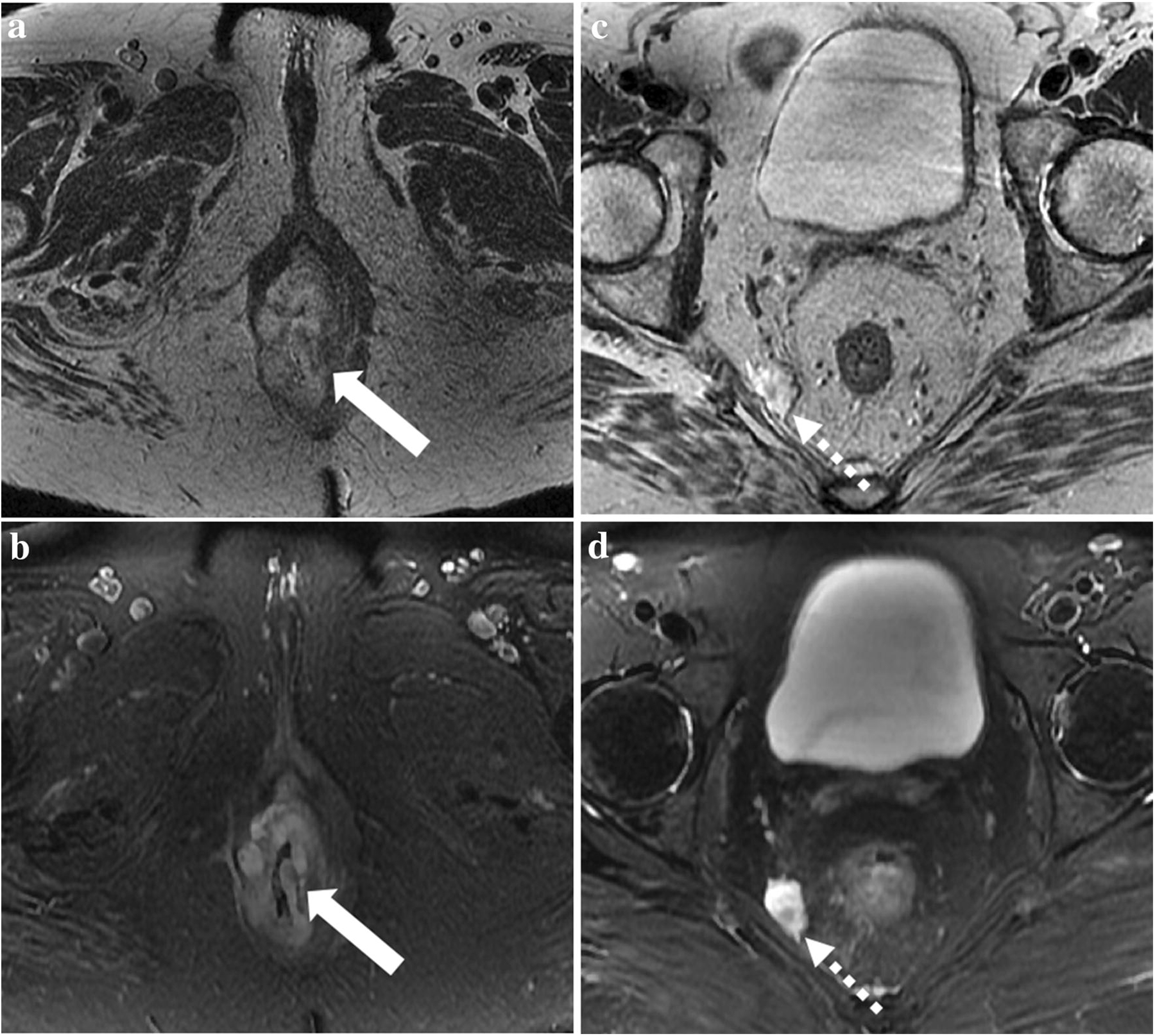
Rectal adenocarcinoma with mucinous components is an uncommon type of rectal cancer with two distinct histologic subtypes: mucinous adenocarcinoma and signet-ring cell carcinoma. Mucin can also be identified as pattern of response after neoadjuvant treatment. On imaging modalities, mucin typically demonstrates high signal intensity on T2-weighted images, low attenuation on computed tomography, and may be negative on 18-fluorodeoxyglucose positron emission tomography. After neoadjuvant CRT, cellular and acellular mucin share similar imaging features, and differentiating them is currently the main challenge faced by radiologists. Radiologists should be aware of pros, cons, and limitations of each imaging modality in the primary staging and restaging to avoid misinterpretation of the radiological findings.

Keywords: Computed tomography; Magnetic resonance imaging; Mucin; Positron emission tomography; Rectal neoplasms.


Conflict of interest statement

Conflicts of interest: The authors declare that they have no conflict of interest.

Figures

Fig. 1
Fig. 1
59-year-old man with mucinous adenocarcinoma of the rectum (arrows) causing bowel obstruction. (A-B) Computed tomography on admission demonstrates a lesion in the upper rectum with heterogeneous enhancement and areas of low attenuation. The lesion causes upstream bowel obstruction (asterisks). Sagittal (C) and axial (D) T2WI shows the typical high signal intensity of the lesion, which was confirmed on fat-suppressed T2WI (E). Contrast-enhanced T1WI demonstrates heterogeneous enhancement that was more evident in the periphery of the tumor and less intense within the central areas of higher mucin content.
Fig. 2
Fig. 2
(A) 42-year-old man with signet-ring cell carcinoma in the high rectum and sigmoid colon. Non-contrast enhanced computed tomography demonstrates a long segment of homogeneous thickening involving the upper rectum and sigmoid colon (arrows), with foci of calcifications (arrowhead). (B) 45-year-old woman with signet-ring cell carcinoma in the rectum. Contrast-enhanced computed tomography shows thickening of the rectum with target appearance (dashed arrow).
Fig. 3
Fig. 3
75-year-old woman with mucinous tumor in the lower rectum. Axial T2WI without fat suppression (A,C) and with fat suppression (B,D) demonstrate the lower rectal tumor with high signal intensity (arrows) infiltrating the sphincter complex (arrowhead) and a tumor deposit close to the mesorectal fascia (dashed arrows). Note that the sequence T2WI with fat suppression demonstrates mucin content with more conspicuity.
Fig. 4
Fig. 4
44-year-old man with Crohn’s disease and perianal fistula who had mucinous rectal adenocarcinoma. 18-FDG PET/MRI on primary staging demonstrates a tumor in the lower rectum (arrow) with predominantly high signal intensity on T2WI (A). There is infiltration of the right internal and external sphincters (arrow). On DWI (B) and ADC map (C) there is a focal area with diffusion restriction on the left which has low signal intensity on T2WI and is avid on FDG-PET (D, dashed arrows), suggesting a more cellular component within the mucin pool. (E) PET/MRI fusion demonstrates that correlation. At 2 o’clock, an intersphincteric fistula with hyper metabolism is also demonstrated (arrowheads).
Fig. 5
Fig. 5
62-year-old woman with mucinous adenocarcinoma of the lower rectum without complete response after chemoradiotherapy on surgical specimen - cellular mucin. (A) Primary staging MRI demonstrates on T2WI the tumor with high signal intensity surrounded by a dark rim, which infiltrates the sphincter complex (arrows). (B) Restaging MRI demonstrates slight reduction in the size of the tumor with similar heterogeneous mucin content (dashed arrow). The patient underwent surgery and residual tumor was detected (cellular mucin).
Fig. 6
Fig. 6
56-year-old man with mucinous rectal adenocarcinoma. Axial (A), sagittal (B) and coronal (C) T2WI demonstrate the tumor in the upper rectum with high signal intensity (arrows) and a positive mesorectal lymph node (dashed arrow), which also demonstrates high signal intensity on T2WI due to mucin content.
Fig. 7
Fig. 7
54-year-old man with mucinous adenocarcinoma of the lower rectum with complete response after chemoradiotherapy on surgical specimen - acellular mucin. (A) Primary staging MRI demonstrates on T2WI the tumor with high signal intensity infiltrating (arrow). (B) Restaging MRI demonstrates no significant change in the tumor, which maintains a heterogeneous mucin content (dashed arrow). The patient underwent surgery and no residual tumor was detected (acellular mucin) and he was classified as having complete pathological response.
Fig. 8
Fig. 8
Two patients with mucinous degeneration post chemoradiotherapy, one with cellular (A,B) and the other with acellular mucin (C,D) detected on surgical specimen. (A,B) 49-year-old man with non-mucinous tumor on primary staging MRI (A, arrow). On restaging MRI after chemoradiotherapy, the tumor demonstrated a significant reduction in size and mucin was detected on the tumor bed (B, dashed arrow); however, on histopathological evaluation cellular mucin was detected. (C,D) 67-year-old man with non-mucinous tumor on initial MRI (C, arrow). Restaging MRI did not show a significant reduction in tumor size, but there was mucinous degeneration of the tumor and a heterogeneous mucin content was demonstrated within the treated area (D, dashed arrow). After surgery, no tumor was detected and the mucin content was acellular.

References

    1. Bosman FT, Carneiro F, Hruban RH, Theise ND. WHO Classification of Tumours of the Digestive System: World Health Organization, International Agency for Research on Cancer, 2010.
    1. Verhulst J, Ferdinande L, Demetter P, Ceelen W. Mucinous subtype as prognostic factor in colorectal cancer: a systematic review and meta-analysis. J Clin Pathol 2012;65(5):381–388. doi: 10.1136/jclinpath-2011-200340 - DOI - PubMed
    1. Nitsche U, Zimmermann A, Späth C, Müller T, Maak M, Schuster T, Slotta-Huspenina J, Käser SA, Michalski CW, Janssen KP, Friess H, Rosenberg R, Bader FG. Mucinous and signet-ring cell colorectal cancers differ from classical adenocarcinomas in tumor biology and prognosis. Ann Surg 2013;258(5):775–782; discussion 782–773. doi: 10.1097/SLA.0b013e3182a69f7e - DOI - PMC - PubMed
    1. Hugen N, Brown G, Glynne-Jones R, de Wilt JH, Nagtegaal ID. Advances in the care of patients with mucinous colorectal cancer. Nat Rev Clin Oncol 2016;13(6):361–369. doi: 10.1038/nrclinonc.2015.140 - DOI - PubMed
    1. Xie L, Villeneuve PJ, Shaw A. Survival of patients diagnosed with either colorectal mucinous or non-mucinous adenocarcinoma: a population-based study in Canada. Int J Oncol 2009;34(4):1109–1115. - PubMed

Publication types

MeSH terms